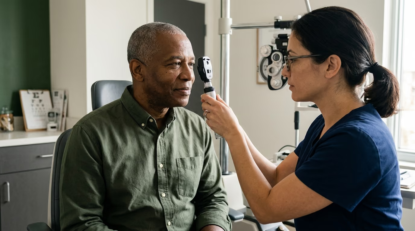

Five core clinical domains explain how military service impacts long term visual processing, physical eye health, and essential healthcare benefits for veterans.





You sit down in front of a computer screen after a poor night of sleep, and the words on the monitor blur into a hazy gray double image. You rub your eyes, blink repeatedly, and assume the eye strain is just fatigue or normal aging. Later that night, driving home on a rain-slicked highway, oncoming headlights create blinding halos that make lane markings disappear completely. You have likely searched for why your vision feels inconsistent despite passing a basic eye chart test, and this guide provides the definitive answers and clinical steps you need.
Vision care for military veterans requires an integrated approach that connects ocular health, brain function, physical trauma history, and daily capability.
Comprehensive eye care goes far beyond updating a lens prescription. It requires evaluating structural eye diseases, assessing neurological visual processing after service-related head trauma, addressing ocular surface damage from harsh environments, and navigating Department of Veterans Affairs healthcare benefits.
Evaluating vision accurately requires understanding that clear eyesight is only one part of visual performance. Eye health spans five distinct domains: optical clarity, structural ocular health, visual processing within the brain, daily functional capability, and consistent clinical access. When any single domain breaks down, overall capability drops.
Optical vision measures how sharply light focuses on the retina across distance, intermediate, and near ranges. Refractive errors like myopia, hyperopia, and astigmatism alter optical clarity, but they represent only mechanical focusing challenges. A veteran may achieve 20/20 distance acuity on a standard wall chart while remaining functionally impaired in complex visual environments.
Ocular health involves the biological condition of the physical eye. This domain includes the transparency of the cornea and lens, the structural integrity of the retina, and the vascular supply supporting the optic nerve. Progressive conditions like glaucoma or diabetic retinopathy often cause severe structural damage before central optical clarity changes.
Visual processing reflects how the brain organizes, interprets, and acts upon visual input. Traumatic brain injury, blast wave exposure, stroke, and systemic illness can disrupt the neural pathways connecting the eyes to the brain. In these cases, the physical structures of the eye may appear normal during a standard examination, yet the individual struggles with spatial orientation, reading tracking, and depth perception.
Functional vision describes how effectively a person uses their sight to complete real-world tasks. It dictates whether a veteran can read small medication labels, step safely off a curb in low light, drive at night, or work on a digital display without debilitating eye strain. Contrast sensitivity, peripheral visual fields, and binocular coordination dictate functional vision far more than basic visual acuity numbers.
Access and continuity of care complete the framework. Veterans must navigate both the Department of Veterans Affairs healthcare system and private community medical networks to secure routine examinations, specialized surgical consultations, low-vision rehabilitation, and necessary corrective eyewear. Understanding how these five domains interact is essential for long-term health, as detailed across our veteran lifestyle and healthcare resources.
Military operational environments expose service members to unique optical and physical hazards. Blast overpressure, penetrating fragments, vehicle rollovers, and blunt-force impacts can cause acute structural injuries and delayed functional problems. Understanding how military trauma affects visual pathways helps veterans identify underlying issues early.
Blast waves generate rapid pressure differentials that compress and distort delicate ocular tissues. Even without a direct penetrating fragment, blast exposure can cause closed-globe trauma, angle recession, retinal tears, and microscopic damage to the optic nerve. VA-supported clinical research indicates that veterans who sustain traumatic brain injuries should undergo comprehensive eye examinations, even if they have no initial visual complaints, to identify hidden structural injuries that could cause secondary glaucoma years later.
Traumatic brain injury frequently damages the neural circuitry controlling binocular vision and oculomotor tracking. Service members returning from deployment may develop convergence insufficiency, where the eyes struggle to turn inward together when reading. This condition causes double vision, visual-motion sensitivity, headaches during screen work, and sudden loss of reading comprehension.
Environmental exposures during deployment also degrade long-term ocular health. Prolonged exposure to burn pits, airborne sand, industrial solvents, toxic chemical agents, and intense ultraviolet radiation damages the ocular surface. These hazards cause chronic inflammatory changes in the conjunctiva and tear-producing glands, leading to persistent discomfort that lingers long after leaving active duty.
Emergency management of acute eye trauma requires strict first-aid discipline. If a penetrating injury or foreign body puncture occurs, you must never rub the eye or rinse it with water. Place a rigid protective shield, such as a cut paper cup, over the eye without applying pressure, and seek emergency medical care immediately. For chemical splash exposures, the protocol reverses: flush the eye immediately with clean water for at least fifteen minutes and transport the individual to an emergency facility.
A routine comprehensive eye examination differs fundamentally from a simple vision screening. Vision screenings, such as those conducted at a motor vehicle office or basic physical, only assess distance acuity using a standard letter chart. Comprehensive examinations evaluate the entire visual system, identifying asymptomatic diseases before permanent sight loss occurs.
VA healthcare benefits cover routine eye examinations and preventive vision testing for all enrolled veterans. These examinations evaluate refractive stability, measure intraocular fluid pressure, assess peripheral visual fields, and examine the internal structures of the eye under dilation. Regular exams serve as a critical component of military health priorities throughout active service and post-service transition.
Diagnostic testing during a comprehensive visit includes several clinical assessments:
Preparation improves the clinical value of an eye appointment. Bring your current prescription glasses, backup frames, sunglasses, and contact lens packaging. Compile a comprehensive medication list, including over-the-counter allergy drops, artificial tears, prescription medications, and nutritional supplements.
Be ready to describe specific visual failures rather than just stating that your vision feels blurry. Explain whether you struggle to read street signs at dusk, miss steps when walking down stairs, experience double vision after thirty minutes on a computer, or feel nauseated in busy grocery store aisles with fluorescent lighting. Documenting specific symptoms gives your optometrist or ophthalmologist the context needed to run targeted functional tests.
Refractive errors represent the most common cause of correctable visual impairment. According to the National Eye Institute, more than 11 million Americans live with uncorrected or correctable visual errors. Roughly half of all adults in the United States lack 20/20 acuity due to untreated refractive conditions.
Myopia, or nearsightedness, occurs when the eye is too long or the cornea is too curved, causing distant objects to focus in front of the retina. Hyperopia, or farsightedness, occurs when the eye is too short, making near focusing difficult. Astigmatism results from an irregularly curved cornea or lens, which distorts light rays across multiple focal points and creates blurred vision at all distances. Presbyopia is the natural, age-related loss of lens flexibility that typically begins in a person's forties, making close-up reading difficult.
Correcting these errors involves tailored lens designs based on daily occupational demands:
Protective eyewear is distinct from everyday cosmetic glasses. Standard prescription lenses can shatter under blunt impact, driving sharp fragments directly into the globe. The American Academy of Ophthalmology recommends that individuals with reduced vision in one eye wear impact-resistant polycarbonate lenses at all times to protect their remaining sight.
Workplace and recreational safety standards demand rigorous compliance. The Occupational Safety and Health Administration enforces strict requirements for eye and face protection in hazardous work environments. Industrial facilities, construction sites, and tactical ranges require lenses stamped with the ANSI Z87.1 safety rating. These frames and lenses withstand high-velocity projectile impacts, resist chemical splashes, and filter hazardous ultraviolet radiation.
Dry eye is a complex, chronic disorder of the tear film and ocular surface affecting millions of adults. It frequently produces burning, stinging, foreign-body sensations, redness, and fluctuating vision that temporarily clears with blinking. For many veterans, environmental exposures during military deployments initiate lasting ocular-surface instability.
The tear film consists of three balanced layers: an inner mucin layer, a middle aqueous layer, and an outer lipid layer produced by the meibomian glands along the eyelid margins. When the meibomian glands become blocked or inflamed, the protective lipid layer breaks down. Without this oil barrier, the watery tear layer evaporates rapidly, leaving sensitive corneal nerves exposed to friction from blinking and dry ambient air.
A common paradox of dry eye is excess tearing. When the ocular surface becomes dehydrated and irritated, corneal nerve endings trigger an emergency reflex from the main lacrimal gland. This flood of reflex tears lacks the necessary oils and mucins, causing watery fluid to run down the cheeks without lubricating the cornea.
Managing dry eye requires a systematic approach rather than relying on over-the-counter redness-relieving drops. Redness-relief drops contain vasoconstrictors that temporarily shrink surface blood vessels, but they do not treat the underlying inflammation and can cause rebound redness.
Veterans managing chronic dry eye symptoms should implement targeted self-care steps:
If basic home therapies fail to provide relief, consult an eye specialist. Prescription treatments may include anti-inflammatory cyclosporine or lifitegrast drops, short-term corticosteroid regimens, or punctal plugs that block tear drainage ducts to preserve natural moisture. Managing ocular inflammation forms a core part of supporting healthy aging and longevity.
Veterans face significant risks from age-related and systemic ocular diseases that can permanently destroy vision if left unmanaged. Understanding the pathology and clinical progression of glaucoma, cataracts, diabetic retinopathy, and macular degeneration ensures veterans seek timely medical intervention.
Glaucoma encompasses a group of progressive eye diseases characterized by damage to the optic nerve head, often linked to elevated intraocular pressure. The National Eye Institute reports that over 2.7 million Americans have glaucoma, with projections reaching 4.3 million by 2030. Glaucoma is often called the silent thief of sight because open-angle glaucoma develops painlessly over many years. Central vision remains sharp while peripheral vision gradually recedes, leaving the individual unaware of the loss until significant damage occurs.
Cataracts involve the gradual clouding of the natural crystalline lens inside the eye. The Centers for Disease Control and Prevention notes that cataracts represent the leading cause of vision loss across the United States, affecting roughly 20.5 million adults aged 40 and older. Symptoms include hazy vision, poor night vision, blinding glare from oncoming headlights, and faded color perception. When cataracts interfere with reading, driving, or work, ophthalmologists perform outpatient surgery to replace the clouded lens with a clear artificial intraocular implant.
Diabetic retinopathy damages the tiny blood vessels that nourish the light-sensitive retina. According to the CDC, diabetic retinopathy is the primary cause of blindness among working-age American adults, affecting roughly 4.1 million individuals. High blood glucose levels weaken capillary walls, leading to microaneurysms, fluid leakage, retinal hemorrhages, and abnormal vessel proliferation. The National Eye Institute notes that one in twelve people with diabetes aged 40 and older develops vision-threatening retinopathy. Maintaining strict control over blood sugar, blood pressure, and lipid levels protects retinal microvasculature.
Age-related macular degeneration specifically attacks the macula, the central region of the retina responsible for sharp, straight-ahead vision. It is the leading cause of permanent close-up visual impairment among adults aged 65 and older. The dry form causes gradual thinning of macular tissues and cellular waste accumulation called drusen. The wet form occurs when abnormal blood vessels leak fluid beneath the retina, causing rapid vision loss. Warning signs include distorted or wavy lines, blind spots in the center of your visual field, and sudden difficulty recognizing faces.
Navigating vision care benefits within the Department of Veterans Affairs requires understanding the administrative distinction between clinical eye care and corrective eyewear coverage. Confusion regarding these rules often prevents eligible veterans from accessing services.
All veterans enrolled in VA healthcare qualify for comprehensive clinical eye care. This benefit covers diagnostic screenings, routine eye examinations, preventive glaucoma evaluations, surgical interventions, and specialty ophthalmology care for diseases like retinal tears or cataracts. Routine vision examinations do not require a service-connected disability rating.
Prescription eyeglasses and medically necessary contact lenses follow stricter eligibility guidelines. The VA provides corrective eyewear without cost only to veterans who meet specific statutory criteria:
When VA medical facilities face scheduling backlogs or lack on-site specialty services, veterans can access private providers through the VA Community Care Network. If appointment wait times exceed statutory thresholds or travel distances to the nearest VA eye clinic are excessive, your VA primary care team can issue a community care referral. This pathway allows you to see a private optometrist or ophthalmologist while the VA covers the cost.
Veterans experiencing severe, uncorrectable vision loss should connect directly with the Visual Impairment Services Team coordinator at their nearest VA medical center. VIST coordinators manage comprehensive low-vision rehabilitation programs, providing adaptive optical devices, high-tech electronic magnifiers, and independent living training to help veterans maintain functional autonomy. Navigating healthcare coverage is an essential component of overall veteran lifestyle and healthcare.
Vision loss impacts every aspect of daily life, from working at a desk to navigating roadways safely. Maintaining personal independence requires implementing practical physical adaptations and understanding motor vehicle safety regulations.
Driving requires acute visual processing, broad peripheral awareness, rapid glare recovery, and strong contrast sensitivity. The National Highway Traffic Safety Administration emphasizes that safe driving relies on detecting low-contrast roadway hazards, spotting pedestrians at dusk, and reading highway markers at high speeds. Many older drivers and veterans with early cataracts or retinal disease struggle significantly under low-illumination conditions.
The NHTSA advises that wearing yellow-tinted or polarized sunglasses during night driving reduces the total light entering the eye, worsening hazard detection. To protect yourself and others on the road:
Home and workspace modifications significantly reduce fall risks and eye strain. Install bright, adjustable task lighting directly over kitchen counters, reading desks, and workbench areas to enhance contrast. Apply high-contrast non-slip tape along the leading edges of stairs, steps, and transitions between flooring materials. Keep primary walking paths clear of clutter, power cords, and loose rugs that create tripping hazards.
In corporate and industrial workplaces, optimize ergonomics to protect vision. Adjust computer displays so the top of the monitor sits just below eye level, roughly an arm's length away. Increase system font sizes, use high-contrast dark-mode display profiles, and reposition screens to eliminate overhead fluorescent glare. For industrial, maintenance, and construction roles, adhere strictly to OSHA eye safety standards, ensuring emergency eyewash stations remain accessible and unblocked at all times.
Clinical errors and delayed treatments often occur when veterans and healthcare providers misinterpret visual symptoms. Reviewing common case patterns helps illustrate how complex vision issues present in real-world scenarios.
A 34-year-old veteran returns from an overseas deployment in a dusty, arid environment, complaining that computer screens blur unpredictably throughout the afternoon. Standard vision testing shows 20/20 distance acuity, leading the veteran to assume the issue is mental fatigue. A comprehensive ocular surface evaluation reveals severe meibomian gland dysfunction and rapid tear break-up time caused by chronic sand and dust exposure. Implementing daily warm compresses, using preservative-free tears, and adjusting screen ergonomics resolves the fluctuating blur completely.
A 28-year-old veteran exposed to an explosive blast during service experiences persistent frontal headaches and reads at half their prior speed. An examination reveals structurally healthy retinas and 20/20 distance acuity, but binocular testing detects convergence insufficiency and impaired saccadic tracking from mild traumatic brain injury. Prescribing base-in prism reading lenses and completing ten weeks of targeted vision therapy restores comfortable tracking and eliminates daily headaches.
A 52-year-old veteran with a history of blunt orbital trauma from a vehicle accident notices no visual changes during daily life. During a routine VA examination, tonometry measures an elevated intraocular pressure of 28 mmHg in the injured eye, and gonioscopy reveals angle recession from the prior trauma. Automated perimetry confirms early peripheral visual field loss from secondary glaucoma. Daily pressure-lowering drops halt disease progression, preserving central vision before irreversible nerve destruction occurs.
A 61-year-old veteran avoids driving at night due to severe starbursts and halos around oncoming headlights, despite reading 20/25 on a standard well-lit eye chart. A slit-lamp examination reveals dense cortical cataract spokes that scatter incoming light across the retina under low-contrast conditions. Outpatient cataract extraction with intraocular lens replacement eliminates the disabling night glare, restoring driving capability.
A 73-year-old veteran with Type 2 diabetes begins missing morning insulin doses and mixing up oral heart medications. Family members assume the errors stem from cognitive decline, but a low-vision assessment reveals central visual distortion caused by diabetic macular edema. Connecting the veteran with the VA VIST coordinator provides large-print talking medication dispensers, high-contrast organizational trays, and retinal anti-VEGF injections that stabilize sight.
When evaluating vision care options, veterans must distinguish established, evidence-based treatments from experimental claims. Relying on unproven therapies can delay necessary medical care and result in permanent vision damage.
Solid scientific evidence supports several core clinical interventions:
In contrast, several widely marketed commercial products lack convincing scientific backing. Over-the-counter blue-light-blocking glasses are heavily marketed to reduce eye fatigue and prevent retinal damage, but major ophthalmological reviews find minimal evidence that blue light from digital displays causes physical eye disease. Behavioral adjustments, blinking, and lubricating drops provide far greater clinical relief for screen strain.
Similarly, commercial eye exercise programs claiming to naturally reverse nearsightedness or cure presbyopia lack peer-reviewed validation. While specialized oculomotor vision therapy successfully treats binocular tracking disorders after brain injuries, it cannot alter the physical shape of the cornea or restore elasticity to an aging crystalline lens. Always verify clinical claims through the BattleVet research library.
Protecting your long-term eye health requires consistent habits and proactive healthcare navigation. Follow this structured checklist to ensure your visual system remains sharp, capable, and protected.
This article is published exclusively for educational and informational purposes and does not constitute formal medical advice, clinical diagnosis, or personalized healthcare treatment plans. Eye anatomy, refractive conditions, post-trauma visual processing deficits, and ocular diseases vary substantially among individuals.
Always consult a board-certified ophthalmologist, licensed optometrist, or qualified VA healthcare provider to evaluate your vision, discuss specific symptoms, and determine appropriate medical interventions. Never ignore professional clinical advice or delay seeking emergency medical treatment because of information read in this guide.
VA healthcare covers routine comprehensive eye examinations, glaucoma screenings, and medical eye treatments for all enrolled veterans. However, the VA covers prescription eyeglasses and contact lenses only for veterans who meet specific eligibility requirements, such as having a compensable service-connected disability, being a former POW or Purple Heart recipient, or having vision loss resulting from conditions like TBI or stroke.
Yes. Explosive blast waves cause rapid pressure fluctuations that can damage internal ocular structures, leading to microscopic retinal tears, angle recession, and secondary glaucoma years later. Blast waves and concussions can also disrupt the neural connections between the brain and eye muscles, causing double vision, reading difficulty, and light sensitivity even when the physical eye appears normal.
When meibomian glands along the eyelid margins fail to produce enough protective oil, the watery tear layer evaporates too quickly. This exposes sensitive corneal nerves to friction and air, triggering an emergency reflex from the lacrimal gland. This sudden flood of reflex tears lacks the necessary lubricating oils, causing tears to spill over your eyelids without hydrating the eye surface.
For chemical splashes, flush the open eye immediately with large amounts of clean water for at least fifteen minutes, then seek emergency medical care. For a puncture, cut, or penetrating trauma, do not rinse the eye, do not rub it, and do not attempt to remove any embedded object. Cover the injured eye with a rigid, protective shield without applying pressure, and proceed immediately to an emergency department.
Follow BattleVet for practical guidance on military and veteran health, strength, recovery, testosterone, sleep and healthy aging. Stay connected for new articles, research backed insights and clear information to help you stay capable for the years ahead.


Build better habits around strength, recovery, sleep, hormones and healthy aging with practical guidance for active military personnel and veterans.
Explore BattleVet