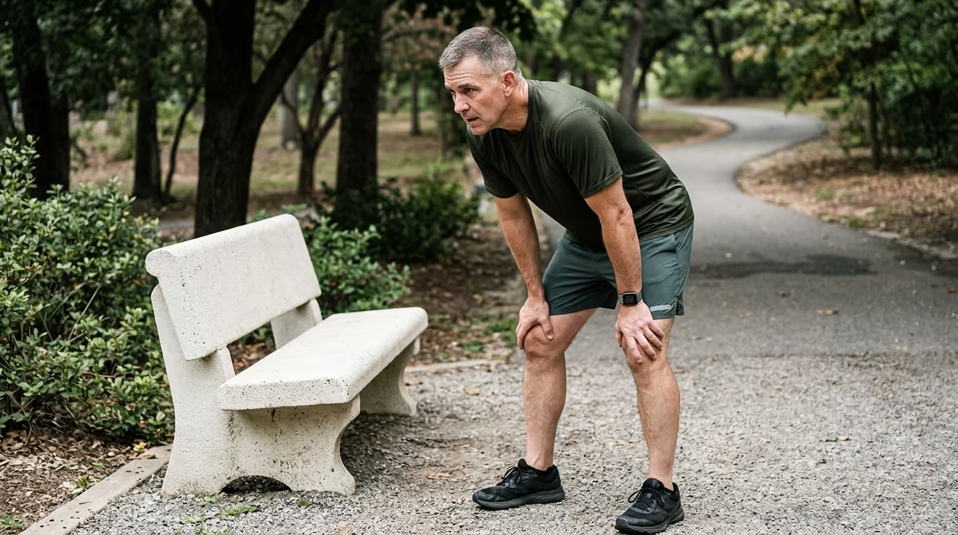

Lower fracture risk and stronger bones reward veterans who combine impact training with key nutritional habits and endocrine health monitoring.





You step off a curb during a morning walk and feel an unexpected, sharp pain in your foot. A follow-up visit reveals a hairline fracture, prompting your doctor to order a bone density scan. Decades of carrying heavy loads, running on paved roads, and pushing through joint discomfort might make you feel physically resilient. However, silent changes in bone remodeling can leave your skeletal system vulnerable long before your first noticeable symptom appears.
Preserving bone density and avoiding fractures after military service requires a deliberate combination of progressive resistance training, adequate nutrition, hormone management, and targeted clinical screening.
Long-term skeletal health is determined by the balance between bone breakdown and rebuilding, which is influenced by mechanical strain, endocrine signals, and nutritional intake. For military veterans, lifetime fracture risk is often elevated due to prior orthopedic trauma, joint wear, long-term medication use, and mobility limitations. Protecting skeletal strength after service requires measuring baseline risk with dual-energy X-ray absorptiometry, performing high-load resistance training, maintaining adequate calcium and vitamin D levels, and addressing secondary causes of bone loss.
Bone is dynamic living tissue that continuously repairs and rebuilds itself through a process called remodeling. Specialized cells called osteoclasts break down old or damaged bone tissue. Meanwhile, cells called osteoblasts lay down new collagen matrix that gradually mineralizes with calcium and phosphate. In early adulthood, bone formation outpaces resorption, leading to peak bone mass around age thirty. After this peak, the balance gradually shifts toward resorption, resulting in a slow loss of bone mineral density over time.
Mechanical loading is the primary physical signal that stimulates osteoblasts to build stronger bone. When your skeleton experiences mechanical strain from lifting weights or absorbing ground impact, specialized sensor cells called osteocytes detect the deformation. These cells send chemical signals that downregulate bone resorption and trigger local bone formation. Conversely, when a limb is unloaded due to injury, cast immobilization, bed rest, or reduced mobility, osteoclasts become more active and bone density drops rapidly.
Veterans face unique physical exposures that influence skeletal remodeling over decades. Heavy operational load carriage, repeated blast exposures, parachute landings, and high-impact training place tremendous cumulative stress on bones and joints. While active mechanical strain can build bone, sustained high-volume training without adequate recovery or energy intake can suppress bone formation. A 2024 study published in military epidemiology literature reported higher fracture risk among veterans than non-veterans, with an unadjusted odds ratio of 1.24 and an adjusted odds ratio of 1.20.
Long-term skeletal health is also tied to functional recovery and physical restoration. When joint injuries, chronic pain, or nerve damage lead to reduced physical activity, the skeleton loses the mechanical loading signals it needs to stay dense. Bone quantity and bone quality both matter. Bone quantity refers to the total mineral mass visible on a scan, while bone quality refers to internal microarchitecture, collagen integrity, and accumulated microscopic damage.
Osteoporosis and low bone mass are silent conditions that rarely cause pain until a fracture occurs. Dual-energy X-ray absorptiometry, commonly known as a DXA scan, is the standard diagnostic tool used to measure bone mineral density. A DXA scan typically evaluates the lumbar spine, the femoral neck of the hip, the total hip, and occasionally the non-dominant forearm. The resulting measurement generates two primary scores: a T-score and a Z-score.
A T-score compares your bone mineral density to the average peak bone density of a healthy young adult of your sex. The World Health Organization defines normal bone density as a T-score of -1.0 or higher. A T-score between -1.0 and -2.5 is classified as low bone mass, historically referred to as osteopenia. A T-score of -2.5 or lower at the spine, femoral neck, or total hip establishes a clinical diagnosis of osteoporosis.
A Z-score compares your bone density to age-matched and sex-matched peers. While T-scores are standard for postmenopausal women and men aged fifty or older, Z-scores are especially valuable for younger adults, active-duty service members, and premenopausal women. A Z-score of -2.0 or lower indicates bone density that is below the expected range for your age group. This finding should always prompt a thorough clinical investigation for secondary causes of bone loss, such as endocrine disorders or malabsorption.
Low bone mass should never be interpreted as complete safety from injury. Because low bone mass is far more prevalent across the population than osteoporosis, the absolute majority of fragility fractures occur in individuals with T-scores between -1.0 and -2.5. A fragility fracture is defined as a fracture resulting from a fall from standing height or less. If a veteran suffers a low-trauma fracture of the hip, spine, wrist, or shoulder after age fifty, clinical guidelines recognize this event as a diagnostic pathway for osteoporosis regardless of the DXA score.
The Fracture Risk Assessment Tool, known as FRAX, estimates a person's ten-year probability of experiencing a major osteoporotic fracture or a hip fracture. Major osteoporotic fractures include clinical fractures of the spine, hip, forearm, and humerus. In the United States, clinical guidelines from the Bone Health and Osteoporosis Foundation identify high risk when the ten-year probability is at least 3 percent for a hip fracture or 20 percent for a major osteoporotic fracture. These numerical thresholds help clinicians determine when prescription medication is warranted alongside lifestyle interventions.
FRAX has practical limitations that veterans should understand. The tool does not fully account for recent multiple fractures, frequent falls, or the severe localized bone loss caused by spinal cord injury or amputation. It also does not capture high-dose medication exposure or severe neuromuscular weakness. A veteran with frequent balance issues or prior blast-related injuries may have a much higher real-world fracture risk than standard FRAX calculations suggest.
Vertebral fracture assessment, performed via specialized DXA software or standard lateral spine radiographs, is a crucial diagnostic step. Vertebral compression fractures are frequently painless or dismissed as simple chronic back strain. Identifying an asymptomatic vertebral collapse provides clear evidence of skeletal fragility and fundamentally changes the clinical management plan.
Physical training is one of the most powerful non-pharmacological tools for building and preserving bone density across the lifespan. Structured physical activity protects the skeleton through two distinct mechanisms. First, it exerts mechanical strain on bones, stimulating osteocytes to reinforce local mineral architecture. Second, it develops neuromuscular strength, power, and coordination, reducing the likelihood of falling.
A complete bone preservation training plan combines progressive resistance training, controlled impact loading, and dynamic balance work. For a broader view of physical training protocols that maintain muscle tissue, review our strength, fitness, and body composition resources.
Resistance exercise must challenge the muscular system with sufficient intensity to trigger skeletal adaptation. Clinical consensus guidelines recommend resistance training two to three days per week, focusing on major compound movement patterns. To stimulate bone formation, sets should generally use loads between 50 percent and 85 percent of your one-repetition maximum, performing 5 to 12 repetitions per set with high technical precision.
The most effective resistance exercises target the muscle attachments around the hip, spine, and upper limbs. These movements include:
Resistance training must be progressive to remain effective. As your body adapts over weeks and months, you must gradually increase the external load, adjust the volume, or modify the movement tempo. Light dumbbells lifted for thirty repetitions without muscular fatigue provide cardiovascular benefits, but they do not create the mechanical strain required for bone remodeling.
For veterans managing chronic joint pain or surgical hardware, resistance training must be customized. Movements can begin with isometric contractions, machine-guided paths, resistance bands, or partial ranges of motion. Working with a physical therapist ensures that joint limitations do not prevent you from applying safe mechanical load to the surrounding bone.
Impact loading generates rapid ground reaction forces that travel up the skeleton, creating dynamic fluid flow within bone tissue that strongly activates osteoblasts. Jumping, hopping, brisk stair climbing, and directional changes stimulate bone adaptation more effectively than non-impact exercises like swimming or cycling. Clinical exercise guidelines suggest aiming for approximately 50 moderate impacts per session on three non-consecutive days each week.
Moderate impact exercises include:
Impact exercise must always be scaled to your current functional capacity and bone status. If you have known spinal compression fractures, multiple fragility fractures, severe joint degeneration, or poor balance, high-impact jumping is contraindicated. In these situations, brisk walking, progressive resistance machines, and supported step-ups provide safe skeletal stimulation without dangerous peak forces.
Because the vast majority of non-vertebral fractures result from falls, fall prevention is just as important as building bone mineral density. A strong skeleton can still break if subjected to the high torsional forces of an awkward fall. Incorporating balance, agility, and lower-body power training into your weekly routine significantly lowers fall incidence.
Effective fall-prevention routines incorporate:
Veterans should evaluate home hazards, optimize lighting, wear supportive footwear with slip-resistant soles, and review sedating medications with their healthcare providers to eliminate preventable fall triggers.
Bone tissue is a complex composite material consisting of a protein-rich collagen matrix reinforced with mineral crystals. Proper nutrition supplies the structural raw materials and regulatory compounds required for continuous remodeling. A diet lacking essential micronutrients or adequate energy forces the body to pull minerals from the skeleton to support critical metabolic functions elsewhere.
Calcium is the primary structural mineral in human bone. The Bone Health and Osteoporosis Foundation recommends a total daily calcium intake of 1,200 milligrams for women older than fifty and men older than seventy. Men aged fifty to seventy should target 1,000 milligrams daily, while younger adults require approximately 1,000 milligrams per day.
Clinical guidelines emphasize obtaining calcium from whole foods whenever possible. Dietary calcium sources are absorbed effectively and carry a lower risk of adverse side effects than high-dose supplements. Excellent dietary calcium sources include:
When dietary intake falls short of daily targets, supplements can fill the gap. Calcium carbonate should be taken with meals because it requires stomach acid for optimal absorption. Calcium citrate can be taken with or without food and is preferable for veterans taking acid-reducing medications or those with low stomach acid. To maximize intestinal absorption, divide supplemental calcium into doses of 500 milligrams or less at a single time.
Excessive calcium supplementation should be avoided. High-dose calcium supplements can cause constipation, increase the risk of kidney stones in susceptible individuals, and interact with medications such as levothyroxine and certain antibiotics.
Vitamin D is an essential hormone-like nutrient that enables calcium absorption in the small intestine. Without sufficient vitamin D, the body absorbs only 10 to 15 percent of dietary calcium, triggering the release of parathyroid hormone, which strips calcium from bones. The National Institutes of Health recommends 600 International Units of vitamin D daily for adults up to age seventy, and 800 International Units daily for adults over seventy. Clinical organizations often recommend 800 to 1,000 International Units daily for older adults or individuals with low bone density.
A large meta-analysis summarized by the National Institutes of Health evaluated eight randomized controlled trials involving 30,970 participants. The researchers found that combined supplementation of calcium and vitamin D in adults older than fifty reduced total fractures by 15 percent and hip fractures by 30 percent. Vitamin D alone without adequate calcium does not consistently reduce fracture rates, emphasizing the need for both nutrients.
Serum 25-hydroxyvitamin D blood tests evaluate your current vitamin D status. Levels below 20 nanograms per milliliter reflect deficiency, while levels between 30 and 50 nanograms per milliliter support optimal skeletal and muscular health. Veterans with limited sun exposure, darker skin pigmentation, gastrointestinal disorders, obesity, or chronic kidney disease often require customized vitamin D3 supplementation protocols supervised by a clinician.
Adequate dietary protein is vital for skeletal structural integrity. Collagen accounts for roughly 90 percent of the organic bone matrix, providing tensile strength and flexibility that prevents bone from becoming brittle. Protein also stimulates the production of insulin-like growth factor 1, a key hormone that promotes bone formation and muscle protein synthesis.
Older adults and veterans should target 1.2 to 1.6 grams of protein per kilogram of body weight daily, distributed evenly across meals. Consuming sufficient protein alongside resistance exercise prevents sarcopenia, the age-related loss of muscle mass and strength that dramatically elevates fall risk.
Chronic energy deficiency and severe caloric restriction undermine bone density. When overall caloric intake is insufficient to support daily energy expenditure, reproductive hormones decline, metabolic rate drops, and bone turnover shifts heavily toward resorption. Maintaining a healthy, stable body weight is a fundamental component of fracture prevention. Low body mass index is an established clinical risk factor for osteoporotic fractures.
Smoking tobacco impairs bone health through multiple pathways. Nicotine and toxic combustion byproducts inhibit osteoblast function, reduce intestinal calcium absorption, accelerate estrogen breakdown, and impair microvascular blood flow to bone tissue. Smoking cessation consistently halts accelerated bone loss and reduces long-term fracture risk.
Heavy alcohol consumption directly damages osteoblasts, disrupts liver metabolism of vitamin D, and impairs balance and motor coordination, drastically increasing fall probability. Limiting alcohol intake to no more than one standard drink per day for women and two for men protects both bone architecture and neuromuscular control.
The skeletal remodeling cycle is tightly regulated by the endocrine system. Fluctuations in systemic sex hormones, thyroid hormones, and parathyroid hormones directly alter osteoclast and osteoblast activity. Understanding your hormonal profile helps identify underlying conditions that accelerate bone loss. For more on navigating these systems, read our analysis on testosterone and hormones in veteran wellness.
Estrogen plays a major protective role in the skeletal systems of both women and men. It suppresses osteoclast activity, slowing the rate of bone resorption and maintaining bone remodeling balance. Following natural or surgical menopause, circulating estrogen levels drop precipitously. This withdrawal leads to a phase of rapid bone loss that can strip away up to 10 to 20 percent of a woman's bone mass within the first five to seven years post-menopause.
Early menopause, defined as menopause occurring before age forty-five, and prolonged premenopausal amenorrhea represent substantial fracture risk factors. Women experiencing early estrogen deficiency should undergo clinical evaluation for early bone density screening and discuss hormonal or non-hormonal bone-protective strategies with their physicians.
In men, testosterone contributes to skeletal health through direct androgen receptor signaling on osteoblasts and through aromatization into estrogen. Primary or secondary hypogonadism leads to accelerated bone resorption and decreased bone formation. While testosterone replacement therapy improves bone mineral density in hypogonadal men, established male osteoporosis with high fracture risk still requires approved anti-fracture medications.
Androgen deprivation therapy, a cornerstone treatment for advanced prostate cancer, profoundly impacts bone density. By suppressing testosterone and downstream estrogen to castrate levels, androgen deprivation therapy triggers rapid bone demineralization, with lumbar spine and hip bone density dropping significantly within the first year of therapy. Clinical guidelines recommend baseline DXA screening, fracture risk assessment, and preventative antiresorptive therapy for veterans undergoing androgen deprivation therapy.
Hyperthyroidism and excessive thyroid hormone replacement therapy accelerate bone remodeling, shortening the cycle and leaving osteoblasts inadequate time to replace resorbed bone matrix. Veterans taking levothyroxine for hypothyroidism should have their thyroid-stimulating hormone levels checked annually to avoid overtreatment that compromises bone density.
Primary hyperparathyroidism occurs when one or more parathyroid glands produce excess parathyroid hormone, pulling calcium out of the bones and into the bloodstream. A routine comprehensive metabolic panel showing elevated serum calcium should always prompt follow-up testing for parathyroid hormone levels. Surgical removal of an overactive parathyroid gland can halt bone loss and restore bone mineral density.
Many common prescription medications alter bone metabolism, promote calcium excretion, or impair bone formation. A regular medication review with your primary care provider is an essential step in identifying secondary osteoporosis risks.
Systemic glucocorticoids, such as prednisone, dexamethasone, and methylprednisolone, are the most common cause of secondary osteoporosis. Glucocorticoids suppress osteoblast differentiation, accelerate osteoblast and osteocyte apoptosis, reduce intestinal calcium absorption, increase renal calcium excretion, and cause muscle wasting that worsens fall risk.
The 2022 American College of Rheumatology guideline addresses adults receiving 2.5 milligrams or more of prednisone daily for more than three months. For these patients, the guideline strongly recommends:
You should never abruptly stop taking prescribed glucocorticoids, as sudden cessation can cause dangerous adrenal insufficiency. Instead, speak with your physician about tapering to the lowest effective dose and adding targeted bone-protection therapies.
Several other classes of long-term prescription medications can influence bone density or fracture risk:
Do not alter or discontinue any prescribed medication on your own. Discuss potential bone risks with your doctor, who can assess alternative drugs, adjust dosages, or order appropriate surveillance scans.
When bone density is very low or fracture risk is elevated, lifestyle and nutritional interventions alone are insufficient to prevent fractures. Modern osteoporosis medications fall into two main categories: antiresorptive agents and anabolic agents.
Antiresorptive medications slow down bone resorption, allowing osteoblasts to maintain or gradually increase mineral density. This class includes:
Anabolic medications directly stimulate osteoblasts to build new, high-quality bone matrix. These daily or monthly injectable medications include teriparatide, abaloparatide, and romosozumab. Anabolic therapies are typically reserved for individuals with severe osteoporosis, very low T-scores, or multiple fragility fractures, and are usually followed by an antiresorptive medication to lock in the newly formed bone.
If you are prescribed denosumab, never delay or skip an injection without medical supervision. Stopping denosumab causes a rapid rebound in bone turnover, resulting in fast bone loss and an increased risk of multiple vertebral fractures within months of discontinuation.
Physical trauma sustained during military service creates complex, localized challenges for skeletal health. Managing bone preservation requires adapting traditional guidelines to accommodate orthopedic hardware, chronic neurological conditions, and altered biomechanics. To read more about navigating the transition to long-term wellness after service, check our veteran lifestyle and healthcare materials.
Spinal cord injury triggers rapid, severe bone demineralization below the level of the neurological lesion. The sudden loss of mechanical loading and neural signaling causes massive osteoclast activation. Veterans with complete spinal cord injuries can lose up to 40 percent of their sublesional bone mineral density within the first two years post-injury.
Bone loss following spinal cord injury is most pronounced around the distal femur and proximal tibia, the bones surrounding the knee joint. Standard lumbar spine and hip DXA scans may show normal or only mildly reduced bone density, failing to capture the extreme fracture vulnerability in the lower legs. Even minor rotational forces during routine wheelchair transfers, bowel care, or spastic muscle spasms can cause low-trauma fractures.
A 2019 study evaluating veterans with spinal cord injury found that completing a DXA scan was strongly associated with receiving appropriate osteoporosis medication, with an odds ratio of 84.03. Furthermore, having a prior fracture was a major clinical predictor of treatment, with an odds ratio of 5.43. Proactive screening and early referral to physical medicine and rehabilitation specialists ensure that wheelchair-dependent veterans receive targeted antiresorptive therapies, safe transfer training, and adaptive standing frame protocols when appropriate.
Limb loss introduces asymmetrical skeletal loading. The intact limb absorbs increased impact forces and carries more total body weight, while the residual limb experiences localized unloading and altered stress distributions from the prosthetic socket. Bone mineral density in the hip on the amputated side frequently declines due to reduced weight-bearing and altered muscular pull.
Joint fusions, reconstruction hardware, and severe chronic joint pain cause compensatory gait adaptations. When a veteran alters their gait to unload an arthritic knee or fused ankle, mechanical stress shifts to the contralateral limb and the lumbar spine. Resistance and impact exercises must be carefully structured around these mechanical asymmetries:
Chronic musculoskeletal pain, sleep apnea, insomnia, and post-traumatic stress disorder represent substantial barriers to bone-preservation strategies. Chronic sleep fragmentation disrupts growth hormone secretion and increases systemic inflammation, accelerating bone loss. Post-traumatic stress and depression can reduce motivation for regular exercise, increase reliance on alcohol or tobacco, and lead to poor nutritional habits.
Treating bone health requires an integrated approach that addresses these foundational health factors. Short, structured exercise sessions, clear daily nutritional routines, peer accountability, and coordinated care between primary care, mental health, and physical therapy make long-term skeletal preservation sustainable.
Making informed decisions about bone health requires distinguishing established clinical standards from preliminary findings. The table below outlines what clinical science has firmly established versus areas that remain under active investigation.
Explore our broader editorial analysis in healthy aging and longevity for more deep dives into evidence-led functional health.
Taking control of your skeletal health requires a systematic, phased plan that addresses medical evaluation, physical training, and nutritional consistency.
Certain skeletal symptoms require urgent medical evaluation rather than self-directed exercise adjustments. Contact your healthcare team promptly if you experience any of the following clinical signs:
A diagnosis of osteopenia, or low bone mass, does not mean your bones are safe from fracture. Low bone mass indicates that your bone density is lower than normal, which raises your overall fracture risk. Because far more people have osteopenia than osteoporosis, the absolute majority of fragility fractures occur in individuals within the osteopenia range. Your total fracture risk depends on your age, fall history, muscle strength, balance, and medications, not just your T-score.
Walking is an excellent activity for cardiovascular fitness, mental health, and general mobility, but walking alone provides insufficient mechanical strain to prevent bone loss. Bone requires dynamic, progressive, and multi-directional loading to stimulate new mineral formation. To protect your skeletal density, you should pair daily walking with progressive resistance training and, if medically safe, controlled impact loading like heel drops, stair climbing, or low-box step-downs.
Men can and do develop osteoporosis. While postmenopausal women experience rapid bone loss due to dropping estrogen levels, men experience a steady decline in bone density as they age. Approximately one in four men over the age of fifty will experience an osteoporotic fracture in their lifetime. For male veterans, risk factors such as prior orthopedic injuries, low testosterone, long-term glucocorticoid use, heavy alcohol consumption, and androgen deprivation therapy make proactive bone screening essential.
You should never abruptly stop taking prescribed medications without direct guidance from your doctor. Discontinuing systemic steroids suddenly can cause life-threatening adrenal crises, and stopping other medications can cause severe rebound symptoms. The correct approach is to review your complete medication list with your physician. Ask if your current dosage can be safely tapered, if alternative medications are available, and whether you should start preventative bone-protection therapies.
This article is provided for educational and informational purposes only and does not constitute medical advice, clinical diagnosis, or treatment recommendations. Individual skeletal health, fracture risk, and medication management require personalized assessment by a qualified physician, endocrinologist, or physical therapist. Always consult your healthcare team before beginning a new exercise program, altering your nutritional intake, or modifying any prescription medication.
Review this guide whenever you receive updated DXA scan results, start a new long-term prescription medication, experience an adult-age fracture, or notice changes in your physical mobility. Maintaining dense bones and functional strength is an ongoing, lifelong commitment that supports your independence and physical capability across every decade after service.
Follow BattleVet for practical guidance on military and veteran health, strength, recovery, testosterone, sleep and healthy aging. Stay connected for new articles, research backed insights and clear information to help you stay capable for the years ahead.


Build better habits around strength, recovery, sleep, hormones and healthy aging with practical guidance for active military personnel and veterans.
Explore BattleVet