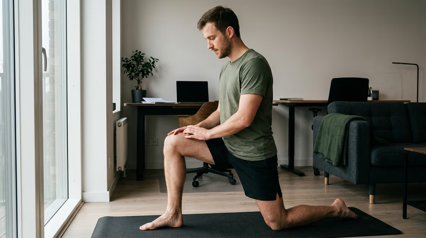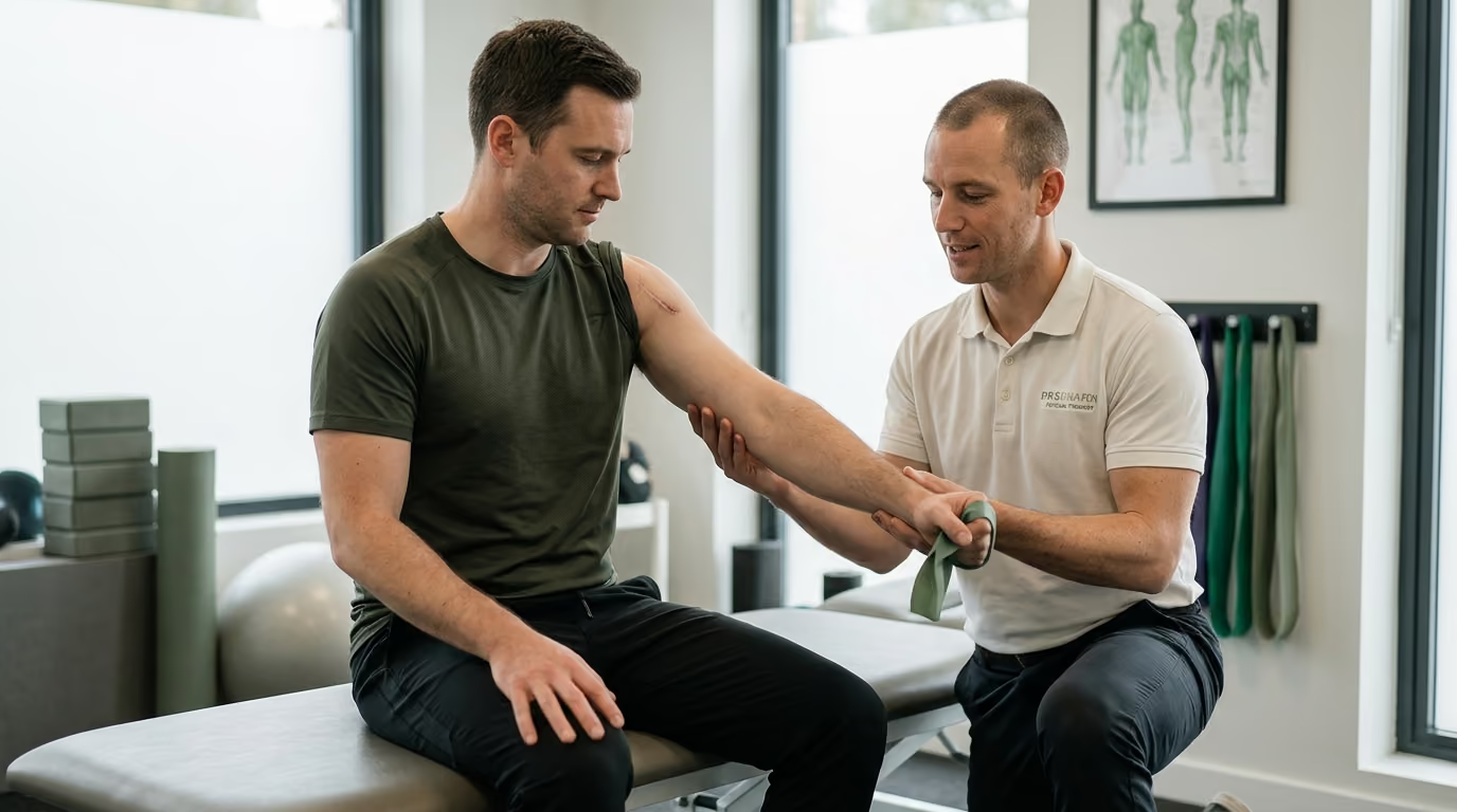

Stepping out of the operating room, patients navigate structured rehabilitation phases from acute healing and early mobility to full athletic performance.





You wake up after surgery with a rigid brace, an aching limb, and an overwhelming list of movement restrictions. Getting out of bed to use the bathroom feels like solving a complex mechanical puzzle. You wonder how much movement is safe, how much pain is normal, and whether pushing harder will speed up your return to normal life or damage the repair.
Orthopedic recovery requires balancing tissue protection with progressive physical loading, ensuring you regain strength and mobility without compromising surgical repair.
Orthopedic surgery alters joint mechanics and temporarily lowers tissue tolerance, meaning recovery is not a passive waiting game. A successful outcome requires a systematic progression through distinct rehabilitation phases. You must protect the surgical repair, systematically restore joint motion, rebuild muscular strength, and progressively retrain real-world movement patterns.
Physical recovery begins with understanding tissue capacity. Every surgical procedure, whether a joint replacement, tendon repair, fracture fixation, or spinal decompression, creates a temporary reduction in what your body can tolerate. Tissue capacity refers to the amount of mechanical load, movement, and stress an anatomical structure can handle before becoming irritated or injured. Rehabilitation demand represents the cumulative physical stress placed on that structure through exercise, walking, household tasks, and daily movement.
When rehabilitation demand exceeds current tissue capacity, the surgical site responds with persistent swelling, increased pain, muscle inhibition, and potential structural breakdown. Conversely, if demand remains too low for too long, secondary complications emerge. Prolonged immobilization leads to joint stiffness, muscle atrophy, bone mineral loss, and cardiovascular deconditioning. The central challenge of orthopedic rehabilitation is finding the precise mechanical stimulus that encourages healing and adaptation without overloading repairing tissues.
Weight-bearing status is one of the most critical mechanical boundaries established by your surgeon. Non-weight-bearing means no body weight passes through the operated limb under any circumstance. Toe-touch or touch-down weight bearing permits the foot to touch the floor purely for balance, but the limb must not support meaningful body weight. Partial weight bearing allows a designated percentage of body weight, often measured using a standard scale or crutch mechanics. Weight bearing as tolerated means loading is guided by pain, swelling, and movement quality, though assistive devices may still be required to prevent a limp.
Range of motion restrictions are equally vital during early recovery. Passive range of motion occurs when an external force, such as a therapist or continuous passive motion machine, moves the joint without muscle contraction from the patient. Active-assisted range of motion involves the patient moving the joint with external support. Active range of motion requires the patient's own muscles to create the joint movement. Each classification places different mechanical stresses across tendons, ligaments, and surgical hardware.
Structured rehabilitation relies on progressive, overlapping phases rather than arbitrary dates on a calendar. While healing timelines provide general boundaries, advancement from one phase to the next should depend on meeting objective criteria.
Prehabilitation takes place before scheduled elective surgeries. The goal is to build physical reserve, optimize movement patterns, and educate the patient on postoperative protocols before entering the operating room.
A comprehensive prehabilitation program targets muscle groups around the affected joint, reinforces core stability, and improves cardiovascular endurance within pain-free boundaries. It also familiarizes the patient with assistive devices, such as walkers or crutches, which reduces anxiety and movement error immediately after surgery. Setting up your living space, planning post-discharge support, and learning proper recovery and sleep habits before the operation substantially improves early confidence.
The primary objective during the immediate postoperative window is protecting the surgical construct while regaining essential functional independence. This phase typically spans the first few days to several weeks, depending on the procedure.
Priorities in this phase include controlling pain, reducing post-surgical edema, preventing deep vein thrombosis, and mastering safe transfers. Basic movements include moving from lying to sitting, standing up safely, walking short distances with an assistive device, and navigating stairs. Isometric muscle contractions, such as ankle pumps and gentle quadriceps sets, are often initiated to prevent complete muscular dormancy without moving the healing joint.
Once acute inflammation subsides and the surgical repair reaches initial stability, rehabilitation shifts toward restoring joint mobility. This phase focuses on active-assisted and active movement within safe parameters.
Clinicians work to eliminate protective muscle guarding and restore normal joint arthrokinetics. For lower-limb surgeries, this stage prioritizes regaining full knee extension or hip extension to restore a normal walking pattern. For upper-limb surgeries, gentle assisted elevation and rotation exercises prevent capsular adhesions. Movement quality remains paramount; forcing a joint through a painful range with compensatory body shifts reinforces poor movement mechanics.
Restoring joint range allows for progressive loading of the surrounding musculature. Muscle weakness after surgery stems from both disuse atrophy and arthrogenic muscle inhibition, a neural process where joint swelling shuts down muscle activation.
Rebuilding strength begins with low-load isometric holds, progressing to closed-chain bodyweight movements, resistance bands, and external loads. Neuromuscular control exercises, including balance drills, single-leg stance, and controlled deceleration, retrain the nervous system to stabilize joints dynamically. Developing a sound foundation in strength and fitness during this phase protects the joint from asymmetrical wear during daily tasks.
Localized joint recovery must be matched with systemic physical conditioning. Weeks of reduced activity degrade aerobic capacity, increase fatigue rates, and impair metabolic health.
Low-impact conditioning modalities can be introduced based on surgical clearance. Stationary cycling, upper-body ergometry, incline treadmill walking, and water-based exercise provide cardiovascular stimulus without high joint impact. Building aerobic capacity enhances blood flow, accelerates systemic recovery, and reduces the fatigue that often leads to movement errors late in the day.
Functional retraining bridges the gap between clinical rehabilitation exercises and daily life requirements. This phase customizes physical training to the specific demands of your home, workplace, and recreational goals.
Exercises in this phase simulate real-world movement patterns. Examples include lifting objects from the floor, stepping over obstacles, carrying loads unilaterally, and performing multi-planar movements. The focus shifts from isolated muscle strength to coordinated, multi-joint movement under varying speeds and loads.
The final phase prepares individuals for physically demanding occupations, heavy manual labor, or competitive athletics. Clearance for this phase is based on objective testing batteries rather than time alone.
Training incorporates higher movement velocities, plyometrics, change-of-direction drills, and sport-specific skill work. Psychological readiness is formally evaluated alongside physical markers. An individual must demonstrate confidence in the repaired limb to prevent compensatory movement patterns during high-intensity tasks.
Modern orthopedic protocols prioritize early, structured mobilization over prolonged bed rest. Extensive clinical literature demonstrates that controlled movement accelerates recovery, reduces hospital stays, and lowers complication rates.
A 2024 systematic review published in clinical literature analyzed 13 studies involving 297,435 patients following hip fracture surgery. The researchers evaluated outcomes between 235,275 patients mobilized early, defined as movement within 24 to 48 hours post-surgery, and 62,160 patients mobilized later. The pooled data demonstrated that early mobilization was associated with lower 30-day mortality, showing an odds ratio of 0.35 with a 95% confidence interval of 0.31 to 0.41. Early mobilization was also associated with a reduced complication rate, showing an odds ratio of 0.43 with a 95% confidence interval of 0.36 to 0.51.
Early movement also influences long-term functional independence. A dedicated study examining older adults after hip fracture repair found that early postoperative mobilization was associated with independent walking at one year, with an odds ratio of 3.79 and a 95% confidence interval of 1.30 to 11.06. However, the study noted that early mobilization was not significantly associated with a full return to pre-injury walking status. This highlights that while early activity promotes independence, overall recovery is still influenced by age, baseline mobility, and underlying health conditions.
Enhanced Recovery After Surgery programs, known as ERAS, incorporate early mobilization within a comprehensive perioperative care pathway. A systematic review evaluating ERAS programs across orthopedic surgery identified a lower overall incidence of postoperative complications in the enhanced recovery cohorts, demonstrating an odds ratio of 0.70 with a 95% confidence interval of 0.64 to 0.78. A broader review of 25 studies involving 16,699 total hip and knee replacement patients similarly linked these multimodal pathways to reduced mortality, lower transfusion rates, and shorter hospital stays.
Evidence regarding prehabilitation shows clear measurable benefits. A 2023 systematic review and meta-analysis evaluated 48 randomized controlled trials involving 3,570 orthopedic patients. The analysis demonstrated that pre-surgical exercise programs significantly improved preoperative functional capacity, muscle strength, and quality of life while reducing back pain. The study confirmed that these functional advantages persisted into the short and medium-term postoperative recovery periods.
Rehabilitation timing requires nuanced application depending on the repaired tissue. In rotator cuff repair, an overview of systematic reviews analyzing 10 systematic reviews and 11 randomized controlled trials compared early mobilization protocols with conservative, delayed mobilization. The researchers found no significant differences between early and conservative rehabilitation regarding functional scores, pain levels, range of motion, or tendon retear rates. However, the evidence was less conclusive for large and massive tendon tears, suggesting that deliberately delaying stress on higher-risk repairs remains a prudent strategy.
Different tissues heal at different rates, and distinct surgical procedures carry unique mechanical vulnerabilities. Rehabilitation protocols must adapt directly to the anatomical region and surgical method.
Total hip and total knee arthroplasty require immediate restoration of functional movement and early weight bearing.
Following total knee replacement, the primary early challenge is overcoming quadriceps inhibition and eliminating knee flexion contractures. Achieving full knee extension is critical for normal gait mechanics and preventing chronic lower back or hip strain. Total hip replacements may require specific positional precautions depending on the surgical approach. Posterior approaches often restrict hip flexion past 90 degrees, internal rotation, and crossing the legs for roughly six weeks to prevent dislocation, whereas anterior approaches may carry fewer movement limitations.
Tendon and ligament reconstructions involve biological healing between soft tissue and bone, which takes considerably longer than joint surface replacements.
Rehabilitation after anterior cruciate ligament reconstruction or rotator cuff repair requires strict adherence to healing timelines. For rotator cuff repairs, active use of the shoulder is restricted during the early weeks, relying on passive range of motion to prevent joint stiffness. The repaired tendon requires progressive, low-tension stimulus to remodel without subjecting the anchor site to disruptive tensile forces. Advancing to active lifting too early can compromise structural integrity.
Surgical repair of fractured bones relies on internal fixation devices such as plates, screws, and intramedullary rods to stabilize bone fragments.
Rehabilitation protocols depend heavily on whether the fixation provides rigid stability or relative stability. Early range of motion of adjacent, uninjured joints is encouraged immediately to prevent stiffness and muscle wasting. However, weight bearing through the fractured bone must follow the surgeon's explicit protocol based on radiographic evidence of bone callus formation. Full loading before adequate biological bridging can lead to hardware loosening or nonunion.
Spinal surgeries, including discectomies, laminectomies, and spinal fusions, require strict movement and posture management.
Early recovery centers on maintaining a neutral spinal alignment, log-rolling when getting in and out of bed, and limiting prolonged seated positions. Spinal fusions require avoiding spinal flexion, rotation, and heavy lifting until bone graft incorporation occurs. Walking serves as the primary early cardiovascular and functional exercise, helping to restore neural mobility and core activation without putting shear stress on the spine. Maintaining broad military health principles such as discipline in posture and deliberate physical movement helps protect spinal integrity long term.
Tracking recovery through objective physical metrics prevents emotional guesswork and ensures training loads remain appropriate for tissue healing.
Pain levels alone do not provide a complete picture of tissue healing, but symptom patterns offer valuable feedback on exercise dosing.
Patients should record pain at rest, pain during physical therapy, and pain the following morning. A mild increase in discomfort during exercise that settles within a few hours is a normal response to progressive loading. However, pain that disrupts sleep, persists into the next morning, or is accompanied by a sudden spike in joint swelling indicates that the previous day's mechanical load exceeded current tissue capacity. Monitoring joint circumference with a tape measure provides an objective metric for swelling management.
Range of motion should be measured systematically using a goniometer or clear functional landmarks.
In addition to joint degrees, clinicians track movement efficiency and gait quality. Distance walked is an incomplete metric if the individual walks with a pronounced limp or excessive trunk lean. Using an assistive device, such as a cane or single crutch, to maintain a symmetrical, pain-free gait is far more beneficial than walking unassisted with poor mechanics. Tools such as the Timed Up and Go test provide standardized data on functional mobility and fall risk.
Strength gains should be assessed through controlled functional movements before progressing to maximal resistance testing.
Clinicians evaluate the ability to perform uncompensated sit-to-stand repetitions, step-ups, and single-leg balance holds. Handheld dynamometry and limb symmetry indices allow therapists to compare strength between the surgical and non-surgical limbs. Progressing to higher-level conditioning, such as running or jumping, generally requires the operated limb to achieve at least 80 to 90 percent of the strength of the uninjured side.
The ultimate benchmark of recovery is the successful return to meaningful daily activities and occupational roles.
Standardized outcome questionnaires, such as the Lower Extremity Functional Scale or Disabilities of the Arm, Shoulder and Hand score, measure practical capabilities. Tracking milestones like showering independently, climbing a flight of stairs step-over-step, driving safely, and completing a full workday provides a clear, holistic picture of physical restoration.
Avoiding common rehabilitation mistakes is just as important as performing your prescribed exercises. Small errors in execution or mindset can create prolonged plateaus or set back structural healing.
Pain typically decreases long before repaired biological tissues regain their full tensile strength. When a surgical site stops hurting, individuals often assume the tissue is fully healed and prematurely resume heavy lifting, impact sports, or high-demand work. Biological remodeling of tendons, ligaments, and bone takes many months. Discontinuing prescribed movement precautions simply because you feel good is a leading cause of secondary injury.
A common misconception among active individuals is that doing more repetitions and more sessions will speed up healing. Bone, cartilage, and tendons require adequate rest intervals to adapt to mechanical loads. Excessive exercise volume leads to chronic joint effusion, persistent tendon irritation, and arthrogenic muscle inhibition. Recovery happens during the rest periods between appropriately dosed training sessions, supported by adequate nutrition and fueling to rebuild tissue.
Assuming you are ready for the next phase of activity simply because a certain number of weeks have passed is an unreliable approach. Tissue healing rates vary based on age, nutritional status, sleep quality, and preexisting health conditions. Advancing to running, lifting, or unrestricted work must be earned by demonstrating adequate joint range of motion, muscle strength, dynamic stability, and absence of swelling under load.
Pushing through a movement with poor mechanics reinforces dysfunctional movement patterns that are difficult to correct later. Walking with a stiff knee, hitching the hip to clear the foot, or shrugging the shoulder to lift the arm places abnormal mechanical stress on adjacent joints. If an exercise cannot be performed with clean form, the load, range, or complexity must be reduced immediately.
While muscle soreness and joint stiffness are normal components of recovery, certain clinical symptoms indicate serious medical complications that require immediate professional attention.
Deep vein thrombosis is a blood clot that forms in the deep venous system, most commonly in the lower leg. The American Academy of Orthopaedic Surgeons identifies calf pain unrelated to the surgical incision, localized tenderness or redness behind or below the knee, and new swelling in the thigh, calf, ankle, or foot as warning signs of a possible clot. If a clot dislodges and travels to the lungs, it becomes a pulmonary embolism, a life-threatening medical emergency. Sudden shortness of breath, sharp chest pain that worsens with deep breathing, dizziness, or coughing up blood require immediate emergency medical care.
Surgical site infections can compromise the repair and lead to serious systemic illness. The Centers for Disease Control and Prevention lists spreading redness around the incision, increased localized heat, progressive swelling, cloudy or foul-smelling wound drainage, and a fever above 100.4 degrees Fahrenheit as hallmark signs of infection. If you notice persistent or expanding redness around the surgical site, contact your surgical team immediately.
Mechanical complications include hardware failure, joint dislocation, or acute tendon re-tear. Warning signs include a sudden, sharp pop accompanied by acute pain, a visible deformity around the joint, an inability to bear any weight through a previously stable limb, or sudden loss of active joint motion. These symptoms warrant immediate cessation of activity and an urgent evaluation by your orthopedic surgeon.
A successful recovery requires converting clinical protocols into a sustainable daily routine. Applying structure to your daily habits ensures consistent progress while safeguarding repairing tissues.
Create a structured daily timetable that distributes your rehabilitation exercises, walking intervals, and rest periods throughout the day. Performing three short, high-quality exercise bouts is far more effective than completing one exhausting session that leaves the joint swollen and painful. Schedule dedicated periods for limb elevation to keep resting edema under control.
Use the 24-hour response rule to gauge whether an exercise progression is appropriate. If your pain increases slightly during an exercise but returns to baseline within two hours, the training dose is well-tolerated. If the joint remains painful, stiff, or visibly swollen the following morning, you have exceeded your current tissue capacity. Reduce the volume, load, or range of motion during the next session until the joint stabilizes.
Do not rush to discard crutches, walkers, or canes. An assistive device reduces joint contact forces and allows you to practice a normal, symmetrical walking pattern. Wean off an assistive device only when you can walk across a room with zero limp, adequate balance, and no compensatory movement. Moving from two crutches to one crutch, or from a walker to a cane, should occur systematically under your physical therapist's guidance.
Recovery demands substantial metabolic energy to rebuild bone, muscle, and collagen. Prioritize adequate dietary protein, micronutrients, and hydration to support tissue remodeling and immune function. Prioritizing quality sleep is equally vital, as growth hormone release and cellular repair peak during deep sleep stages. Engaging with structured veteran life resources can also assist in managing stress and maintaining focus throughout an extended rehabilitation period.
Use this practical checklist to guide your physical rehabilitation this week:
Normal post-exercise pain feels like muscular fatigue, mild stretching discomfort, or a temporary dull ache around the joint that subsides within two hours after stopping. Dangerous pain presents as sharp, stabbing sensations during movement, sudden joint instability, pain that escalates through the night, or discomfort accompanied by rapid joint swelling. If resting pain worsens or if you experience mechanical clicking paired with acute pain, pause your program and consult your physical therapist or surgeon.
Post-surgical swelling can persist for six to twelve months, particularly in lower-extremity joints like the knee and ankle due to gravity and daily standing demands. Low-grade inflammation continues as internal tissues remodel and adapt to progressive physical loading. However, if swelling increases suddenly, is accompanied by localized heat and redness, or severely restricts your established range of motion, it should be evaluated by a healthcare professional to rule out infection or overload.
You cannot force biological tissue healing to occur faster by simply adding more exercise volume. Tendons, ligaments, and bones remodel in response to measured mechanical stress, but they require adequate recovery intervals to synthesize new collagen and strengthen structural matrices. Doing excessive therapy often leads to chronic inflammation, joint effusion, and muscle inhibition, which delays long-term functional recovery rather than accelerating it.
You are ready to transition away from an assistive device when you can bear full weight through the operated limb without pain, maintain single-leg balance, and walk with a completely normal, symmetrical gait. If you discard your device too early and walk with a noticeable limp, you reinforce dysfunctional mechanics and place excessive stress on your lower back, hips, and opposite leg. Always consult your physical therapist before discontinuing an assistive device.
This article is for educational purposes only and should not be used as a substitute for professional medical advice, diagnosis, or treatment. Orthopedic surgical procedures, anatomical repairs, and individual healing timelines vary significantly from person to person. Always consult your orthopedic surgeon, physical therapist, or qualified healthcare professional regarding your specific surgical restrictions, weight-bearing precautions, rehabilitation protocols, and symptom management. Never ignore professional medical instructions or delay seeking care based on information you have read online.
Follow BattleVet for practical guidance on military and veteran health, strength, recovery, testosterone, sleep and healthy aging. Stay connected for new articles, research backed insights and clear information to help you stay capable for the years ahead.


Build better habits around strength, recovery, sleep, hormones and healthy aging with practical guidance for active military personnel and veterans.
Explore BattleVet