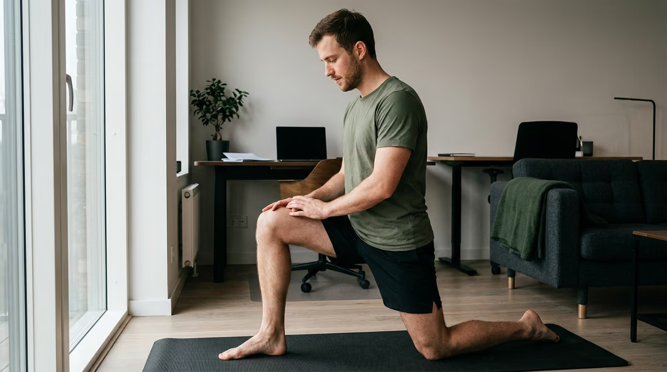

Rest might seem safe, but progressive mechanical loading drives the cellular mechanotransduction required to rebuild durable tendon stiffness and strength.





You step out of bed, place your weight onto the floor, and feel an immediate, sharp ache in the back of your heel or below your kneecap. You rested the joint for two weeks, took anti-inflammatory pills, and avoided hard training, but the stiffness returned the moment you jogged across the street. You search online for answers, wondering why tendon pain seems to defy standard recovery timelines. This guide provides a definitive, evidence-led breakdown of how tendons heal, why rest alone fails, and how structured loading rebuilds long-term tissue capacity.
Restoring tendon function requires progressively loading the tissue to increase its mechanical capacity, rather than waiting for pain to vanish through complete rest.
Tendinopathy is a clinical condition characterized by pain, reduced function, and diminished load tolerance. It is not simply an inflammatory event, nor is it identical to an acute muscle tear. When a tendon is overloaded repeatedly beyond its current capacity, its cellular matrix becomes disorganized. True recovery occurs when you expose the tendon to controlled, progressive mechanical strain. This strain stimulates cellular adaptation, aligns collagen fibers, and restores the tissue's ability to store and release elastic energy under high physical demand.
Tendons connect muscle to bone, acting as biological springs and force transmitters. They must withstand massive tensile forces while maintaining joint stability. To understand why tendon rehabilitation requires patience, one must examine the unique cellular architecture of tendon tissue.
Tendons consist predominantly of parallel type I collagen fibers embedded within an extracellular ground substance composed of proteoglycans, glycoproteins, and water. Tenocytes, the resident specialized fibroblast cells, sit between these collagen bundles. Tenocytes monitor the mechanical environment and regulate matrix synthesis and degradation.
Unlike skeletal muscle, healthy tendon tissue has a relatively low metabolic rate and limited vascular perfusion. While this low metabolic demand allows tendons to sustain tension for long periods without ischemic necrosis, it also results in a slow rate of tissue turnover and structural remodeling.
Tendon adaptation relies on a cellular process called mechanotransduction. When mechanical load stretches a tendon, physical force transmits through the extracellular matrix to cell-surface receptors known as integrins and focal adhesion complexes. This physical trigger activates intracellular signaling cascades, including the MAPK/ERK, PI3K/Akt, and YAP/TAZ pathways.
These biochemical signals instruct tenocytes to produce new collagen proteins, adjust enzyme activity, and reorganize the extracellular matrix. Research shows that mechanical strain stimulates both anabolic protein synthesis and catabolic matrix breakdown. The net biological outcome depends directly on the magnitude, frequency, and duration of the applied load.
When an acute structural tendon injury or surgical repair occurs, healing follows three overlapping phases:
This initial stage lasts roughly one to seven days. Platelets, neutrophils, and macrophages migrate to the injury site to clear cellular debris. Growth factors and inflammatory cytokines recruit progenitor cells, forming an early, fragile fibrin clot.
Beginning within days and lasting several weeks, this phase involves rapid cellular division and matrix synthesis. Tenocytes produce large quantities of disorganized type III collagen and ground substance, establishing a temporary repair bridge with low mechanical strength.
Starting around six to eight weeks post-injury, this critical phase continues for many months. Type III collagen is gradually replaced by stronger, highly oriented type I collagen. The extracellular matrix consolidates, cellularity decreases, and collagen fibers align along the lines of mechanical stress.
Because full structural remodeling requires months of progressive physical demand, reducing pain does not mean the tendon has returned to full mechanical capability.
Designing an effective rehabilitation plan requires an understanding of how tendons respond to physical forces. Tendon capacity is multidimensional and cannot be measured solely by how much weight a person can lift.
Tendon capacity represents the total volume, velocity, frequency, and magnitude of mechanical load a tendon can endure without developing structural failure or persistent symptom flares. A person may possess enough capacity to walk five miles on flat pavement, yet lack the capacity to sprint fifty yards or jump off a curb.
Tendon behavior is governed by several core biomechanical parameters:
Stiffness describes the amount of force required to deform or elongate a tendon by a specific distance. Greater stiffness allows for more rapid and efficient force transmission between the contracting muscle and the skeleton.
Elastic modulus represents the intrinsic material stiffness of the tendon tissue, independent of its physical dimensions. Resistance training improves tendon stiffness primarily by increasing the elastic modulus of the tissue rather than solely increasing tendon thickness.
This refers to the overall thickness of the tendon. While cross-sectional area can increase over long periods of heavy training, structural hypertrophy occurs slowly compared to changes in tissue density and material stiffness.
Strain is the percentage of elongation relative to the resting length of the tendon. In human Achilles tendons, research indicates that mechanical strain between 4.5 percent and 6.5 percent provides the optimal stimulus for tenocyte mechanotransduction and collagen remodeling. Strains below this window may fail to stimulate sufficient adaptation, while excessive strains without adequate recovery can provoke reactive tissue overload.
Achieving a productive mechanical strain generally requires loading intensities above 70 percent of a person's maximum voluntary contraction. However, external resistance must always be balanced against joint angles and movement velocity.
Tendon load consists of tensile force and compressive force. Tensile load occurs when the muscle contracts and pulls along the longitudinal axis of the tendon. Compressive load occurs when the tendon wraps around a bony prominence, such as the Achilles tendon compressing against the calcaneus during deep dorsiflexion, or the proximal hamstring tendon compressing against the ischial tuberosity during deep hip flexion. Insertional tendinopathies are particularly sensitive to compressive forces, requiring exercises that limit extreme joint ranges during early rehabilitation.
Rehabilitation exercise selection should match the irritability of the tendon and the specific mechanical deficit being addressed. Rather than relying on a single exercise philosophy, modern rehabilitation utilizes a continuum of distinct loading modalities.
Isometric contractions generate internal muscle-tendon tension without visible joint movement. This modality serves as a valuable entry point when dynamic joint movement is painful or during an acute symptom flare.
A landmark crossover trial by Ebonie Rio and colleagues evaluated the immediate response to isometric loading in athletes with patellar tendinopathy. Five sets of 45-second isometric knee extensions at roughly 70 percent of maximum voluntary effort produced an immediate reduction in tendon pain. This analgesic effect lasted for at least 45 minutes, alongside a reduction in cortical muscle inhibition.
Isometrics offer specific advantages:
Despite these benefits, systematic reviews indicate that isometric exercise is not universally superior to dynamic exercise for long-term clinical outcomes. Isometrics should be viewed as an entry point and symptom-management tool rather than a standalone cure.
Eccentric loading emphasizes the controlled lengthening phase of a muscle-tendon unit under load. In 1998, Hakan Alfredson popularized an eccentric calf-raise protocol for chronic midportion Achilles tendinopathy, prescribing 180 repetitions per day across 12 weeks.
Early trials reported high success rates, establishing eccentric training as a clinical standard. Eccentric contractions subject the tendon to high tensile forces while minimizing energetic cost.
However, subsequent research has modified how eccentric training is applied:
Heavy-Slow Resistance, often abbreviated as HSR, utilizes heavy loads, typically between 70 percent and 85 percent of a one-rep maximum, performed with a deliberate, slow tempo. A standard tempo uses a three-second lifting phase and a three-second lowering phase.
A classic study by Kongsgaard and colleagues compared 12 weeks of HSR training against eccentric decline squats and corticosteroid injections for patellar tendinopathy. The HSR group achieved significant symptom improvement, increased fibril density, and enhanced collagen turnover. At the six-month follow-up, both the HSR and eccentric groups maintained their improvements, while patients receiving corticosteroid injections experienced clinical relapse. The HSR group also demonstrated the highest long-term patient satisfaction.
HSR offers measurable load progression, trains both concentric and eccentric phases, and stimulates favorable structural adaptations within the tendon matrix. Because of the slow movement speed, peak impact forces are eliminated, making it a reliable foundation for building tissue capacity.
Tendons function like elastic springs during locomotion. When running, jumping, or changing direction, the tendon rapidly absorbs, stores, and releases kinetic energy.
Slow strength exercises build basic force capacity, but they do not prepare a tendon for high-velocity energy storage. Plyometric exercises train the rate of force development and the stretch-shortening cycle.
Activities in this category include:
Energy-storage loading places high peak forces on the tendon in fractions of a second. This training must be introduced gradually after the individual demonstrates tolerance to slow, heavy strength loading.
Because structural tendon remodeling occurs over months, pain cannot serve as an on-off switch for activity. Experiencing mild discomfort during exercise does not indicate ongoing tissue destruction. Rehabilitation requires an objective symptom-monitoring framework.
Tendon response is evaluated over a 24-hour cycle rather than judging an exercise purely by how it feels during the set. Tendons often feel warm and comfortable during physical activity, only to become stiff and irritable the following morning.
Clinical guidelines suggest that pain up to 5 on a 0-to-10 scale during or immediately after exercise can be acceptable. However, this is conditional on two strict rules:
Morning stiffness and initial weight-bearing pain upon getting out of bed serve as the primary indicator of tendon tolerance.
Use this three-part guide to monitor training adjustments:
A common mistake in tendon rehabilitation is the boom-bust cycle. When symptoms improve, individuals often jump directly back into intense running, lifting, or field training. The unadapted tendon quickly flares up, forcing the individual to stop all activity.
Complete rest reduces pain temporarily, but it also causes surrounding muscle atrophy and further decreases tendon stiffness. When activity is resumed, the tendon possesses even less capacity than before, triggering an immediate relapse.
Guidance for midportion Achilles tendinopathy emphasizes maintaining modified, tolerable physical activity while executing rehabilitation. Total rest should be avoided for non-acute tendinopathy.
To resolve a symptom flare, modify the highest-cost mechanical load first. High-speed running, jumping, and deep compressive loading should be reduced before eliminating basic resistance training. This approach preserves systemic fitness and muscular strength while protecting the recovering tendon.
Every tendon operates under distinct anatomical and mechanical demands. Rehabilitation programs must be tailored to the specific tendon involved and its functional role.
The Achilles tendon experiences tensile loads up to six to eight times body weight during running and jumping. It is susceptible to midportion pathology, located two to six centimeters above the heel bone, and insertional pathology at the calcaneus.
Often called jumper's knee, patellar tendinopathy involves localized pain at the inferior pole of the patella. It is provoked by deep knee flexion, rapid deceleration, and explosive jumping.
This condition involves pain at the ischial tuberosity, often aggravated by sitting on hard surfaces, lunging, sprinting, and deep hip flexion.
The supraspinatus and infraspinatus tendons stabilize the humeral head against the glenoid fossa. They are vulnerable to compressive and tensile overload during repeated overhead tasks.
Individuals managing systemic recovery factors can read our dedicated guides on recovery and sleep and strength, fitness, and body composition for supporting strategies.
Rebuilding tendon capacity requires an eight-step progression that transitions from early symptom modulation to full physical demand.
Confirm the affected tendon and differentiate chronic tendinopathy from acute tears, bursitis, or nerve entrapments. Determine whether symptoms are tensile-dominant, compression-sensitive, or both.
Select an exercise variation that can be performed with minimal or acceptable discomfort. This may involve isometric holds, bilateral movements, or exercises performed through a reduced range of motion.
Track symptom response during the session, two hours post-exercise, and the following morning upon waking. Use morning stiffness as the primary guide for adjusting volume and intensity.
Transition from static holds to dynamic, slow isotonic lifting. Utilize both concentric and eccentric phases with a three-to-four-second tempo in each direction. Progress external resistance systematically over 8 to 12 weeks.
Gradually increase joint range of motion, exposing the tendon to combined tensile and compressive loading. Monitor for any delayed irritability as joint angles deepen.
Once the tendon tolerates heavy slow loads without flare-ups, introduce fast-velocity movements. Begin with low-amplitude hops and skips, gradually progressing to high-impact jumping and explosive bounding.
Structure training to reflect the specific demands of your sport, occupation, or daily physical routine. A service member or athlete should gradually reintroduce loaded rucking, sprint intervals, and agility drills.
Tendon capacity diminishes when mechanical stimuli are removed. Maintain at least one weekly session of heavy-slow resistance training and periodic dynamic exposures to preserve tissue stiffness and remodeling adaptations.
Those navigating long-term physical resilience during and after active service can reference our overview on military health guidance.
Tendon recovery is frequently hindered by common misconceptions and diagnostic errors. Avoiding these pitfalls ensures efficient rehabilitation.
Tendinopathy involves cell disorganization and matrix remodeling without sudden structural discontinuity. An acute tear or complete rupture presents with sudden, sharp trauma, an audible pop, visible swelling, bruising, or immediate loss of motor function. Acute structural ruptures require immediate clinical evaluation and distinct orthopedic protocols.
Ultrasound and MRI scans frequently show tendon thickening, structural disorganization, and increased fluid in individuals who have zero physical symptoms. Conversely, a person can experience severe tendon pain while imaging appears relatively normal. Rehabilitation progress must be guided by pain behavior, strength metrics, and movement quality, not by repeat imaging scans.
Passive modalities such as massage, ice, ultrasound, shockwave therapy, and dry needling may provide temporary pain relief. However, they do not provide the mechanical strain required for cellular mechanotransduction and structural matrix remodeling. Passive therapies cannot substitute for progressive active loading.
Corticosteroid injections can produce short-term pain reduction by suppressing local biochemical pathways. However, clinical trials consistently demonstrate high relapse rates, impaired long-term collagen synthesis, and reduced tendon structural integrity following steroid injections. They do not increase long-term tendon capacity.
When progressing a program, adjust only one variable at a time. Increasing weight, repetition volume, movement speed, and training frequency simultaneously makes it impossible to identify which factor caused a symptom flare-up.
To begin restoring tendon capacity this week, implement the following practical steps:
This article is published for educational and informational purposes only. It is not intended to provide medical advice, diagnosis, or treatment. Tendon pain can result from various conditions, including acute tears, inflammatory arthritis, metabolic disorders, and nerve compression. Always consult a qualified healthcare professional, physical therapist, or orthopedic physician before initiating a rehabilitation program or making changes to your physical routine.
Tendons are viscoelastic tissues with relatively low sensory nerve density within the central matrix. During exercise, warmth, increased local blood flow, and endorphin release often mask tendon strain. Over the following 12 to 24 hours, cellular biochemical turnover and localized fluid shifts increase within the tendon sheath, creating noticeable stiffness and discomfort when you first bear weight the next morning.
While diagnostic ultrasound or MRI scans may continue to show regions of structural thickness or matrix disorganization, the surrounding healthy tendon tissue can undergo significant adaptation. Research shows that healthy portions of the tendon hypertrophy and increase their material stiffness to compensate for disorganized zones. A person can achieve full, pain-free athletic performance even if repeat imaging continues to show minor structural changes.
In most cases of non-acute midportion tendinopathy, complete rest is counterproductive. You can generally continue modified running or lifting provided that pain during the activity remains mild (below 5 on a 10-point scale), symptoms return to baseline within 24 hours, and morning stiffness does not progressively worsen over the course of the week. Ballistic speeds, hill sprints, and deep compressive ranges may need temporary modification.
You are typically ready to introduce energy-storage drills when you can perform heavy-slow resistance training at near-maximal loads without pain flares, have symmetrical single-leg strength, and report zero next-day morning stiffness from standard strength sessions. Reintroduce plyometrics gradually, beginning with low-amplitude double-leg hops before progressing to single-leg bounding, deceleration drills, and maximum-speed running.
Follow BattleVet for practical guidance on military and veteran health, strength, recovery, testosterone, sleep and healthy aging. Stay connected for new articles, research backed insights and clear information to help you stay capable for the years ahead.


Build better habits around strength, recovery, sleep, hormones and healthy aging with practical guidance for active military personnel and veterans.
Explore BattleVet