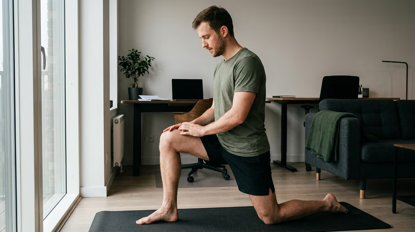

A sudden sharp pull during a sprint requires accurate injury assessment, modern acute care, and progressive loading to rebuild full muscle capacity.





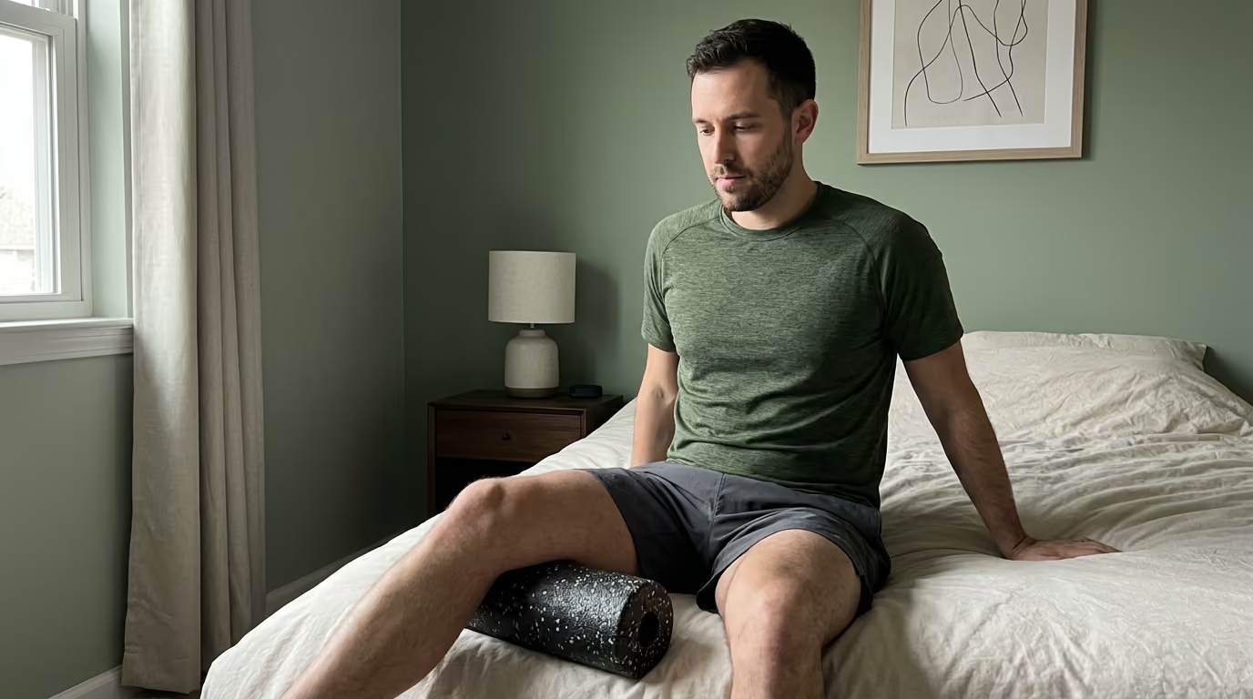
You step out of bed after a strenuous sprint session or field exercise, and a sudden sharp pull in your hamstring or calf brings you to a halt. Walking downstairs feels precarious, your sleep was interrupted by a dull ache, and normal physical training seems weeks away. A muscle strain is an acute overload injury involving muscle fibers, the musculotendinous junction, or the surrounding aponeurotic tissue. It is not simply a tight knot or temporary fatigue that can be rubbed away with a foam roller. True rehabilitation requires a systematic progression from tissue protection to full capacity rebuilding.
A muscle strain heals through structured, progressive mechanical loading that rebuilds the tissue's capacity to handle high force, long lengths, and rapid velocities, rather than through passive rest alone.
To recover from a muscle strain, you must move beyond pain relief and restore the structural and functional capacity of the damaged muscle. Early management focuses on symptom control and gentle movement without excessive inflammation suppression. As healing begins, you progress through isometric loading, full-range eccentric strengthening, lengthened-position loading, and gradual running progressions. Return to full activity requires passing objective strength and speed criteria rather than relying on arbitrary timelines or the absence of resting pain.
A muscle strain occurs when the mechanical demand placed on a muscle exceeds its structural capacity. This overload happens most frequently during forceful lengthening contractions, rapid acceleration, jumping, or sudden changes of direction. The injury can occur within the muscle belly itself, at the musculotendinous junction where muscle transitions into tendon, or across the aponeurosis. Differentiating a muscle strain from referred nerve pain, direct contusions, or complete tendon avulsions is essential for planning recovery.
Clinical sports medicine distinguishes between functional and structural muscle injuries. A functional muscle injury involves pain, muscle tightness, and performance deficits without identifiable macroscopic fiber disruption on diagnostic imaging. These functional disorders often respond quickly to active recovery and load management.
A structural muscle injury involves clear anatomical disruption of muscle fibers, connective tissue, or tendon architecture. Structural injuries present with localized swelling, hematoma formation, and measurable strength loss. Because structural disruptions require cellular remodeling and collagen realignment, they demand a more cautious, phased rehabilitation timeline.
The presence of aponeurotic or tendon involvement changes the recovery timeline significantly. Research on calf muscle strain injuries demonstrates that severe aponeurotic disruption extends return-to-play timelines considerably. In one clinical cohort, athletes with aponeurotic disruption required an average of 31 days to return, compared to roughly 19 days for those with isolated muscle belly strains.
Traditional clinical classifications categorize muscle strains into three distinct grades based on symptom severity and tissue damage:
While these grades provide a common descriptive language, they are imperfect predictors of individual recovery time. A grade alone cannot dictate the exact date an individual is ready for high-intensity activity. Factors such as the precise anatomical location of the tear, the degree of tendon involvement, and the individual's baseline capacity dictate the actual rehabilitation trajectory.
An accurate assessment identifies the damaged tissue, establishes baseline function, and rules out severe injuries requiring surgical consultation. Clinicians evaluate the history of the injury alongside targeted physical tests to understand the mechanism and extent of damage.
The evaluation begins with a detailed history of the injury event. Key factors include the exact movement performed, whether a sudden pop or snap was felt, the immediate ability to bear weight, and any previous history of injury to that muscle group.
Physical examination compares the injured limb directly to the uninjured side. The clinician assesses resting posture, gait abnormalities, localized tenderness, and palpable defects in the muscle tissue. Active range of motion and passive stretch tolerance are measured systematically. Resisted muscle contractions are tested across multiple joint angles to evaluate force production and pain response.
A single physical test is rarely definitive on its own. For example, acute hamstring injuries typically present with an altered walking stride, focal tenderness along the biceps femoris or semitendinosus, pain with resisted knee flexion, and pain during straight-leg hip flexion.
Magnetic resonance imaging and diagnostic ultrasound are the primary imaging tools used to evaluate muscle injuries. Imaging helps confirm the anatomical location, measure the length and cross-sectional area of the tear, and evaluate deep connective tissue involvement. Ultrasound provides a dynamic, accessible evaluation of superficial tissues, while magnetic resonance imaging offers superior visualization of deep musculature and intrameduscular tendons.
Diagnostic imaging has clear limitations. A scan shows anatomical structure, but it does not measure functional capacity, fatigue resistance, or psychological confidence. Research confirms that imaging findings do not always correlate perfectly with readiness to return to strenuous activity. An athlete may display persistent imaging abnormalities long after functional capacity has returned, or show normal scans while still experiencing referred neural pain.
Certain clinical signs indicate a severe injury or an underlying medical emergency that requires prompt specialist evaluation:
Traditional acute management relied heavily on the RICE protocol, which emphasized rest, ice, compression, and elevation. Modern rehabilitation science has evolved toward active management models that optimize tissue regeneration.
The British Journal of Sports Medicine introduced the PEACE and LOVE framework to guide soft tissue recovery from the acute stage through long-term conditioning. This evidence-based approach balances initial protection with early progressive loading.
During the first several days, the PEACE protocol guides immediate care:
Once the acute inflammatory phase subsides, the LOVE protocol governs ongoing recovery:
Inflammation is not a disease process that must be eliminated immediately. It is the biological mechanism by which the body clears cellular debris and delivers growth factors to damaged muscle fibers. Blunting this cascade with high-dose non-steroidal anti-inflammatory drugs or prolonged ice applications can delay the early phases of tissue regeneration.
Medication decisions should always be balanced against pain severity and individual clinical needs. If severe pain disrupts sleep or prevents basic daily mobility, short-term pain relief strategies may be appropriate under a doctor's guidance. However, routine suppression of mild inflammation is no longer recommended as a primary rehabilitation strategy. Readers can review comprehensive insights on tissue recovery within our recovery and physical restoration resources.
Rehabilitation requires a practical symptom-monitoring framework rather than an extreme rule of zero pain. Working with low-level, stable discomfort during controlled exercises is often safe and productive.
To ensure exercise dosages remain within safe biological limits, monitor the 24-hour response:
Successful rehabilitation follows a phased progression designed to systematically rebuild tissue capacity. Progression must be based on objective functional criteria rather than arbitrary calendar dates.
The primary goal of Phase 1 is to settle acute symptoms while restoring baseline mobility. Complete bed rest is avoided because prolonged inactivity causes rapid muscle atrophy, reduced neuromuscular coordination, and disorganized scar tissue formation.
Early movement begins with gentle active range of motion within pain-free boundaries. Low-intensity isometric contractions are introduced to stimulate muscle protein synthesis without subjecting the healing tissue to tensile lengthening stress. For a hamstring strain, this involves gentle double-leg bridge holds or prone knee-flexion isometrics. For a calf injury, seated double-leg calf raises provide low-load activation. For a groin strain, gentle ball squeezes between the knees activate the adductors safely. General cardiovascular fitness is maintained through upper-body training, stationary cycling, or pool walking.
Phase 2 begins once walking is comfortable and low-load isometric contractions are pain-free. The objective is to restore concentric and eccentric strength across stable, predictable movement patterns.
Strength exercises progress from bilateral movements to unilateral variations. Load and volume are increased systematically:
Muscle strength is highly angle-specific. A muscle may generate adequate torque in a shortened position while remaining vulnerable when stretched. Phase 2 introduces varied joint angles to build strength throughout a larger portion of the muscle's operational range. For more detailed training strategies, explore our strength, fitness, and body composition resources.
Most acute muscle strains occur when the muscle is forced to produce high levels of force while rapidly lengthening. Phase 3 targets this specific vulnerability by loading the muscle in extended, elongated positions.
Eccentric training stimulates the addition of sarcomeres in series, which lengthens muscle fascicles and improves the tissue's ability to absorb high-velocity tensile strain. Research from the Academy of Orthopaedic Physical Therapy highlights eccentric training at long muscle lengths as a cornerstone of modern muscle rehabilitation.
Key exercises in this phase include:
These movements must be phased in cautiously. Unaccustomed eccentric training causes significant structural stress and delayed-onset muscle soreness, requiring adequate recovery periods between exposures.
Running places dynamic, high-strain demands on lower-extremity musculature that cannot be duplicated with gym-based resistance training alone. Returning to running requires a structured progression of velocity, acceleration, and deceleration.
A standard running progression advances through distinct stages:
Running volume and intensity should not be increased during the same training session. Athletes must monitor their next-day symptom response after every increase in running speed.
Maximal sprinting and explosive power represent the final frontier of soft tissue rehabilitation. True maximum velocity sprinting creates the highest recorded eccentric forces on the hamstring and calf complexes.
Power reintroduction begins with low-amplitude plyometric drills. Double-leg pogo hops, jump-rope variations, and box jumps teach the muscle-tendon unit to store and release elastic energy efficiently. Once bilateral plyometrics are tolerated, single-leg hopping, bounding drills, and reactive change-of-direction exercises are introduced.
Maximal-speed sprinting is then integrated under structured conditions. Running at 100 percent effort requires full neural recruitment, extreme stretch-shortening cycles, and rapid pelvic stabilization. Athletes should perform high-speed sprints only when completely warmed up and free of residual fatigue.
While general tissue healing principles apply universally, distinct muscle groups possess unique biomechanical roles and recovery requirements.
The hamstring complex, comprising the biceps femoris, semitendinosus, and semimembranosus, is the most frequently strained muscle group in running and field sports. The long head of the biceps femoris is particularly vulnerable during the late swing phase of sprinting, when it must decelerate the rapidly extending knee while preparing for ground strike.
Hamstring rehabilitation requires heavy emphasis on eccentric strength, hip-extension power, and trunk stabilization. Pelvic control exercises, such as dead bugs and single-leg Romanian deadlifts, prevent excessive anterior pelvic tilt, which can place excess passive stretch on the proximal hamstring attachment.
Prior hamstring injury is the single largest predictor of future injury. Meta-analyses indicate that an individual with a previous hamstring strain has nearly three times the risk of sustaining another strain compared to an uninjured peer. Rebuilding eccentric hamstring strength and maintaining regular exposures to maximal sprint velocity are vital to counteract this elevated baseline risk.
The calf complex consists of two primary muscles with distinct anatomical profiles:
Calf rehabilitation must target both structures independently. Straight-knee calf raises emphasize the gastrocnemius, while bent-knee calf raises bias the soleus.
Calf strains often involve the central aponeurosis or the myotendinous junction, which can require prolonged remodeling periods. Recovery timelines reported across sports medicine literature range widely, from 2 to 12 weeks, depending on the depth and tissue type involved. Rehabilitation must progress methodically from slow, heavy strength training to rapid elastic loading and ankle stiffness drills.
Groin strains most commonly affect the adductor longus muscle during cutting, kicking, lateral pivoting, or forceful hip abduction. These injuries are prevalent in field sports that demand rapid changes of direction.
The Copenhagen adduction exercise has emerged as a gold-standard movement in groin rehabilitation and prevention. Research published in sports physical therapy literature demonstrates that progressive Copenhagen adduction training significantly increases eccentric hip adduction strength, reduces groin pain, and improves self-reported functional capacity.
Groin rehabilitation must also address pelvic stability, abdominal strength, and hip rotational mobility. Progressions begin with short-lever Copenhagen holds supported at the knee, advancing to long-lever variations supported at the ankle, before transitioning into dynamic lateral lunges, change-of-direction drills, and kicking protocols.
Quadriceps strains predominantly involve the rectus femoris, the only biarticular component of the anterior thigh. The rectus femoris is vulnerable during kicking, explosive acceleration, and rapid hip extension combined with knee flexion.
Rehabilitation focuses on progressive knee-extension strength, reverse Nordics, and quad-dominant deceleration drills. Less common strains, such as those affecting the abdominals or upper-body musculature, follow the same foundational rules: isolate the biomechanical action of the damaged tissue, restore strength across its full working length, and systematically reintroduce speed and sport-specific demands.
One of the most common mistakes in soft tissue management is clearing an individual based solely on the absence of pain during daily life. True readiness requires meeting objective physical benchmarks that confirm the tissue can withstand competition-level stresses.
Resting pain typically disappears long before structural healing is complete and functional capacity is fully restored. When an individual rests for several weeks, the injured muscle becomes quiet, but it also undergoes disuse atrophy and loses neuromuscular coordination. Returning to full duty or athletic competition simply because walking is painless frequently leads to immediate reinjury.
Rehabilitation must continue through a continuum of milestones:
Objective physical testing provides measurable data to guide return-to-sport decisions:
Limb symmetry indices must be interpreted carefully. If an individual has rested completely, the uninjured limb may have deconditioned as well. Comparing an injured limb to an undertrained contralateral limb can produce false confidence.
Physical performance batteries must bridge the gap between clinical testing and unrestricted activity:
Relying on arbitrary timelines, such as clearing all Grade 2 strains at exactly four weeks, ignores biological variability. Tissue healing rates depend on vascularity, nutrition, sleep quality, age, and individual training history. Some athletes require longer periods of tissue remodeling, while others progress rapidly through movement milestones. Rehabilitation should always be criterion-based rather than calendar-based. Readers can access further foundational knowledge in our military and veteran health resources.
Muscle strains have notoriously high reinjury rates. In professional sports, recurrence rates for hamstring strains are reported between 12 and 48 percent, with the vast majority of reinjuries occurring within the first two months of returning to full activity.
Epidemiological research has identified key risk factors associated with primary and recurrent muscle strains:
The most effective prevention strategy is maintaining regular exposure to the exact forces that caused the initial injury. Eliminating sprinting or heavy loading out of fear creates a fragile, deconditioned muscle.
To protect against recurrent strains, athletes must incorporate micro-doses of high-velocity running and heavy eccentric loading into their weekly maintenance routines:
Many muscle strains occur near the end of training sessions or competitions when systemic fatigue degrades movement coordination. Fatigue impairs neuromuscular timing, leading to improper pelvic positioning and delayed muscle activation.
Rehabilitation and ongoing training must develop local muscular endurance alongside maximal strength. Managing acute-to-chronic workload ratios ensures that total weekly running volume and sprint distance increase gradually, avoiding dramatic spikes that outpace the body's adaptive capacity. For broader strategies on maintaining long-term physical capability, visit our healthy aging articles.
Reviewing clinical case patterns helps illustrate how progressive rehabilitation principles are applied to real-world scenarios.
A recreational runner feels a sudden pull in the posterior thigh during a sprint workout. Walking is slightly uncomfortable, but there is no large hematoma or palpable defect. Early management utilizes the PEACE protocol, incorporating gentle prone knee flexion isometrics and stationary bike riding. By day five, walking is normal, and rehabilitation advances to single-leg bridges, dumbbell Romanian deadlifts, and slider curls. By week two, the runner initiates straight-line jogging and controlled strides, progressing to 90 percent accelerations by week three. Return to unrestricted sprinting is permitted at four weeks after demonstrating symmetrical eccentric strength and completing repeated maximal sprints without symptoms.
A tactical athlete experiences sudden calf pain while pushing a heavy sled. Ultrasound reveals a Grade 2 strain of the medial gastrocnemius with aponeurotic involvement. Because aponeurotic injuries require longer remodeling periods, rehabilitation is paced cautiously. The athlete begins with seated soleus isometrics and non-weight-bearing ankle mobility before progressing to double-leg standing raises at two weeks. Single-leg heel drops and low-amplitude pogo hops are introduced at four weeks. Running progressions begin at week six, with return to full field training achieved at nine weeks following complete resolution of focal tenderness and symmetrical single-leg hop testing.
A soccer player feels sharp groin discomfort during a rapid cut. Straight-line jogging is tolerable, but lateral movement and kicking provoke pain. Rehabilitation prioritizes isometric adductor squeezes, advancing quickly to short-lever Copenhagen holds and lateral band walks. By week three, long-lever Copenhagen planks and lateral lunges are performed under heavy resistance. Dynamic cutting drills and progressive kicking protocols are introduced during week four. The athlete returns to match play at five weeks after passing full-intensity change-of-direction tests with zero next-day symptom aggravation.
An individual presents with ongoing posterior thigh aching during running, yet diagnostic magnetic resonance imaging shows no muscle fiber disruption or hematoma. A detailed physical examination identifies lumbar spine stiffness, positive slump testing, and altered neural dynamics along the sciatic nerve. Rather than treating an isolated muscle strain, rehabilitation shifts toward lumbar spine mobilization, core stabilization, and neural gliding exercises. Symptoms resolve as spinal and neural mechanics improve, allowing a safe return to running.
An athlete sustains a minor hamstring strain, rests completely for three weeks until walking is painless, and immediately joins a full-speed practice session. During the first maximum sprint, the muscle tears again at the exact same location. This recurrent injury occurred because complete rest eliminated resting symptoms without restoring eccentric strength, fascicle length, or high-speed tolerance. The second recovery requires a full eight-week criterion-based rehabilitation program, incorporating heavy eccentric loading and graded sprint exposures to build durable capacity. Readers interested in recovery management can read further in our recovery and sleep articles.
Clear communication regarding scientific evidence allows individuals to make informed decisions about their recovery. Sports medicine literature provides varying degrees of certainty regarding different rehabilitation modalities.
Substantial clinical evidence supports the following principles:
Other areas of soft tissue management remain under active investigation with mixed or preliminary evidence:
Recovering from a muscle strain requires proactive management, consistent execution, and objective monitoring. Follow these practical steps to initiate a structured rehabilitation plan:
This educational guide is provided for informational purposes only and does not constitute individualized medical advice, diagnosis, or treatment. Muscle strains can vary widely in severity, and symptoms may overlap with other musculoskeletal or vascular conditions. Always consult a qualified physician, physical therapist, or healthcare provider to receive an accurate diagnosis and individualized rehabilitation plan tailored to your specific physical condition.
Recovery timelines depend on the severity of the strain, the specific muscle involved, and the presence of tendon or aponeurotic damage. Mild Grade 1 strains often resolve within two to four weeks with proper active rehabilitation. Moderate Grade 2 structural tears typically require six to twelve weeks to fully restore strength, lengthened-position capacity, and sprinting tolerance. Severe Grade 3 ruptures or injuries involving central tendons may require three to six months of extensive rehabilitation or surgical intervention.
No, aggressive stretching should be avoided during the acute phase of a muscle strain. When muscle fibers are torn, pulling them into a deep stretch places excessive mechanical tension on the fragile healing tissue, which can worsen the tear and delay repair. Gentle active range of motion within pain-free boundaries is recommended early on. Stretching should only be reintroduced gradually in later phases, integrated with active strength training through full range of motion.
Persistent or recurring pain often indicates that the muscle healed with reduced structural capacity, shortened fascicle length, or residual weakness. If rehabilitation ended as soon as resting pain disappeared, the tissue may remain unable to handle high force, rapid lengthening, or sprint velocities. Persistent symptoms can also stem from referred neural tension, joint restrictions in adjacent areas, or unaddressed pelvic and trunk mechanics that continue to overload the muscle.
While ice has traditionally been used to numb acute pain, modern sports medicine cautions against excessive, prolonged icing. Ice causes localized vasoconstriction, which can slow the delivery of cellular repair factors and delay the natural inflammatory process essential for early healing. If pain is severe, brief ice application for 10 to 15 minutes may provide temporary analgesic relief, but it should not be relied upon as a primary healing modality.
Follow BattleVet for practical guidance on military and veteran health, strength, recovery, testosterone, sleep and healthy aging. Stay connected for new articles, research backed insights and clear information to help you stay capable for the years ahead.


Build better habits around strength, recovery, sleep, hormones and healthy aging with practical guidance for active military personnel and veterans.
Explore BattleVet