
Stepping out of a cast or walking boot requires navigating five progressive rehabilitation phases to restore mobility, strength, and daily performance.





You wake up to the quiet friction of a rigid walking boot resting against your sheets. Getting out of bed is no longer an automatic reflex, requiring calculated adjustments to keep weight off an injured limb. Even simple morning tasks, like walking to the kitchen or carrying a cup of coffee, suddenly require careful planning and physical effort. Fracture recovery is not merely a waiting game for bone to fuse, but an active process of rebuilding strength, mobility, and confidence.
Quick Take: Full recovery from a bone fracture requires matching your physical activity to the biological stability of the bone, advancing load progressively rather than relying on time or X-rays alone.
Fracture recovery is the phased restoration of structural bone integrity, joint mobility, muscular strength, and task-specific capacity following a skeletal injury. True functional return goes beyond biological bone union. It requires restoring neuromuscular control, rebuilding load tolerance, and safely meeting the physical demands of daily life, work, and physical training.
Bone healing and functional recovery describe two related processes that move at different speeds. Fracture union refers strictly to the biological and mechanical repair of the bone tissue itself. Functional recovery represents your ability to use the limb to walk, climb stairs, lift loads, work, and train without dangerous compensation.
Medical literature shows that radiographic union often lags behind clinical recovery by two to six weeks. A fracture may feel stable during an examination before an X-ray shows complete bone consolidation. Conversely, a bone can show solid structural union on imaging while the surrounding muscles remain weak, the nearby joints stay stiff, and balance remains impaired.
Modern orthopedic rehabilitation evaluates recovery across several distinct levels:
Relying solely on an X-ray to determine physical capacity creates blind spots. An individual might have clear structural union on an image but lack the calf strength or ankle mobility needed to walk down stairs safely. Another person might feel zero pain during daily activities yet lack the impact tolerance required for running or jumping.
Recovery requires evaluating what your body can actually do alongside structural healing. This balanced approach protects the healing bone while rebuilding physical capability. For practical strategies on rebuilding tissue tolerance and managing training fatigue, review our resources on recovery and physical restoration.
Rehabilitation protocols depend directly on the mechanical stability of the fracture site. Stability describes the ability of the broken bone segments to resist displacement under physiological loads. Clinicians evaluate several structural variables to determine how much protection an injury requires.
Fracture location plays a major role in determining mechanical stability. A break in the shaft of a long bone behaves differently than a break that crosses into a joint surface. Articular fractures demand strict anatomical alignment because minor surface irregularities can lead to accelerated joint wear and post-traumatic arthritis.
The pattern of the break also dictates how the injury handles physical load:
Weight-bearing orders must be precise and clearly explained. The British Orthopaedic Association emphasizes that weight-bearing instructions should be reviewed at every follow-up visit and tailored to the patient. These terms define the exact mechanical load permitted through the limb:
A common misunderstanding is that weight-bearing as tolerated means pushing through severe discomfort. In clinical practice, it means applying controlled load that does not cause severe pain, abnormal limping, or delayed swelling.
Orthopedic rehabilitation has shifted away from prolonged, strict immobilization toward early, controlled mechanical loading. Bone is dynamic tissue that responds positively to physiological stress, a principle known as Wolff's Law. Controlled compressive forces stimulate cellular activity, encourage callus mineralization, and reduce the rapid loss of muscle mass and bone density.
Clinical research supports early loading in specific, stable injuries. A 2024 meta-analysis of randomized controlled trials examined early weight-bearing after surgically treated ankle fractures. The researchers found that early weight-bearing significantly improved functional scores at six, twelve, and 24 weeks compared to delayed loading. Patients returned to their pre-injury daily activities an average of 2.74 time units sooner without any significant increase in surgical complications.
Similar results appeared in the UK multicenter WAX trial, a large randomized study evaluating early versus delayed weight-bearing after ankle fracture surgery. At four months, patients allowed to bear weight early showed higher functional scores on the Olerud-Molander ankle scale. The researchers concluded that early weight-bearing was clinically non-inferior, safe, and cost-effective.
Clinical guidelines also endorse early mobilization for older adults recovering from hip fracture repair. Guidance linked to the American Physical Therapy Association recommends immediate weight-bearing as tolerated whenever surgical fixation quality permits. Early mobilization reduces post-operative complications, preserves baseline independence, and lowers mortality risks in older populations.
However, early loading must not be applied universally without considering the specific fracture type. Clinical trials supporting early weight-bearing focus on stable constructs with rigid internal fixation or stable, non-displaced fracture patterns like isolated Weber B fibular fractures. Unstable joint fractures, complex tibial plateau breaks, severe open fractures, and compromised surgical repairs still require structured protection to avoid hardware failure or displacement.
Research from the National Institute for Health and Care Excellence (NICE) highlights that early movement should protect healing tissues while preventing muscular deconditioning. The priority is matching the timing and magnitude of mechanical load to the biological stage of the fracture. You can learn more about structured athletic development and tissue loading through our training and performance guidance.
Fracture rehabilitation follows five progressive phases based on tissue healing, movement quality, and functional capacity rather than arbitrary calendar dates.
The first phase begins immediately following the injury or surgical repair. The primary objectives are protecting bone alignment, managing pain, minimizing edema, and ensuring safe movement during basic daily tasks.
Rehabilitation focuses on maintaining safe mobility without overloading the fracture site. For lower-limb injuries, this involves mastering crutches, walkers, or knee scooters for transfers, stairs, and personal care. Upper-limb injuries require learning single-handed self-care strategies and supporting the limb in a sling or brace.
Movement during this phase preserves range of motion in unaffected adjacent joints:
Phase 1 establishes safe daily habits and protects healing tissues from sudden, unexpected loads.
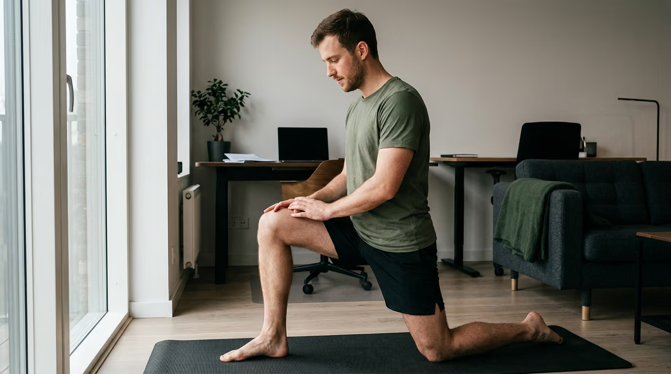
Phase 2 begins once the medical team confirms that initial tissue stability allows active or passive joint motion. The goal is reversing joint stiffness, mobilizing healing soft tissues, and reducing protective muscle guarding.
Active-assisted and active range of motion exercises are introduced systematically. The emphasis during this stage is on smooth, controlled movement rather than aggressive stretching. Forceful stretching can stress healing bone, irritate surrounding tendons, or disrupt surgical hardware.
Key priorities during Phase 2 include:
Movement quality takes priority over volume. Moving cleanly through an available range of motion produces better long-term functional mechanics than forcing a stiff joint past its biological limit.
Phase 3 introduces progressive resistance, higher mechanical loading, and functional strength exercises. This phase begins when clinical examination and follow-up imaging confirm sufficient bone stability to tolerate increased force.
Muscular atrophy develops quickly during periods of casting or non-weight-bearing protection. Rebuilding muscle strength requires progressive overload that challenges the motor units above and below the fracture site.
Rehabilitation protocols during Phase 3 emphasize closed-chain exercises and stability training:
Strength recovery should be measured through functional performance rather than manual muscle tests alone. Tracking single-leg stance time, repeated chair rises, and walking speed provides clear feedback on functional capacity. Explore our resources on strength and physical conditioning for practical frameworks on progressive resistance training.
Phase 4 bridges standard rehabilitation exercises with the specific physical demands of your occupation, sport, or daily routine. The training environment shifts from isolated joint exercises to complex movement patterns and fatigue management.
Conditioning must reflect the actual mechanical stresses you will encounter outside the clinic:
NICE guidelines recommend using functional tasks like lateral stepping, stair climbing, and loaded carries rather than relying solely on gym machines. Phase 4 prepares your musculoskeletal system to absorb unpredictable physical stresses safely.
The final phase manages the safe transition back to unrestricted occupational, athletic, and recreational activities. Clearance is determined by functional testing and movement symmetry rather than arbitrary time frames.
Returning to high-demand activities works best as a graded progression rather than an immediate return to full duty:
Systematic reviews show that return-to-sport timelines vary considerably across different body regions. A simple finger fracture may allow competition within three to four weeks, whereas an operative ankle or tibia fracture often requires several months of conditioning. Structured progression ensures that cardiovascular fitness, dynamic balance, and muscular power match the demands of your activity.
Bone healing is a complex biological sequence that requires cellular signaling, adequate vascular supply, and sufficient nutritional building blocks. Several modifiable and non-modifiable factors can slow healing or lead to delayed union or nonunion.
A major systematic review evaluating surgically managed traumatic fractures identified key clinical risk factors for nonunion. The analysis reported that open fractures increased nonunion risk by 2.6 times, active smoking increased risk by 1.7 times, diabetes increased risk by 1.6 times, and surgical site infection increased risk by 7.0 times. Obesity was also associated with a 1.5 times increase in nonunion risk.
The American Academy of Orthopaedic Surgeons (AAOS) highlights several biological and lifestyle factors that directly affect bone healing:
Addressing modifiable recovery factors supports the body's natural bone repair mechanisms. Maintaining adequate daily protein intake, optimizing dietary calcium and vitamin D, managing sleep quality, and eliminating nicotine products significantly improve the physiological environment for bone regeneration.
Fractures resulting from low-energy mechanisms or simple falls warrant an evaluation of underlying bone mineral density. The National Osteoporosis Foundation recommends assessing bone health and secondary fracture risk after adult fractures. Identifying conditions like osteopenia or osteoporosis allows for proactive medical management that protects your long-term skeletal health. You can read more about preserving bone density and mobility through our healthy aging and longevity resources.
Navigating fracture rehabilitation requires balancing structural protection with active physical restoration. Avoiding common clinical pitfalls prevents unnecessary delays, setbacks, and secondary injuries.
Healing timelines provided at an initial visit are general estimates rather than guarantees. Assuming that six weeks on a calendar equals full structural strength is a common mistake. Biological healing rates vary based on fracture type, fixation method, age, and overall health. Clearance to advance load must always come from clinical examination and diagnostic findings.
Pain often subsides well before a fracture has fully consolidated. Feeling good during basic movements does not mean the bone can tolerate high-impact loads or aggressive lifting. Advancing weight-bearing or dropping protective boots without clinical clearance can overload healing bone and loosen internal fixation hardware.
While protecting the injured bone is essential, whole-body bed rest is counterproductive. Prolonged immobilization leads to cardiovascular deconditioning, rapid muscle wasting, joint contractures, and reduced neuromuscular coordination. Safe, permitted movement for the rest of your body maintains conditioning, supports mental health, and speeds up subsequent rehabilitation phases.
A common rehabilitation error is increasing walking distance, removing a walking boot, climbing stairs, and returning to the gym on the same day. Changing multiple variables at once makes it difficult to pinpoint what caused any subsequent flare-up in pain or swelling. A structured approach changes one variable at a time and monitors the body's response over 24 to 48 hours.
Restoring full joint movement is an important early milestone, but it does not represent complete recovery. A joint can move freely through its normal range while lacking the strength, power, or stability to absorb load safely. True functional recovery requires rebuilding muscular force, single-leg balance, and movement control under fatigue.
Using crutches, walkers, or canes places heavy physical demands on the hands, wrists, shoulders, and upper back. Patients with underlying shoulder impingement, wrist arthritis, or limited upper-body strength often struggle to follow non-weight-bearing restrictions safely. Upper-limb capacity must be evaluated when selecting mobility aids and planning early rehabilitation.
Returning to work based solely on a job title frequently leads to setbacks. A mechanic, construction worker, or nurse faces dynamic physical demands that differ sharply from desk work. A safe return-to-work plan analyzes specific occupational tasks, such as ladder climbing, awkward lifting, and standing duration, systematically rebuilding capacity for each requirement.
A 2025 systematic review of work-related musculoskeletal injuries found that the mean time to return to work was approximately 102 days across multiple studies. This research highlights that occupational recovery is a multi-month process requiring structured conditioning. For broader context on managing physical recoveries and navigating health systems, visit our health and wellness resources.
Different fracture patterns, anatomical locations, and patient populations present distinct challenges that require customized rehabilitation strategies.
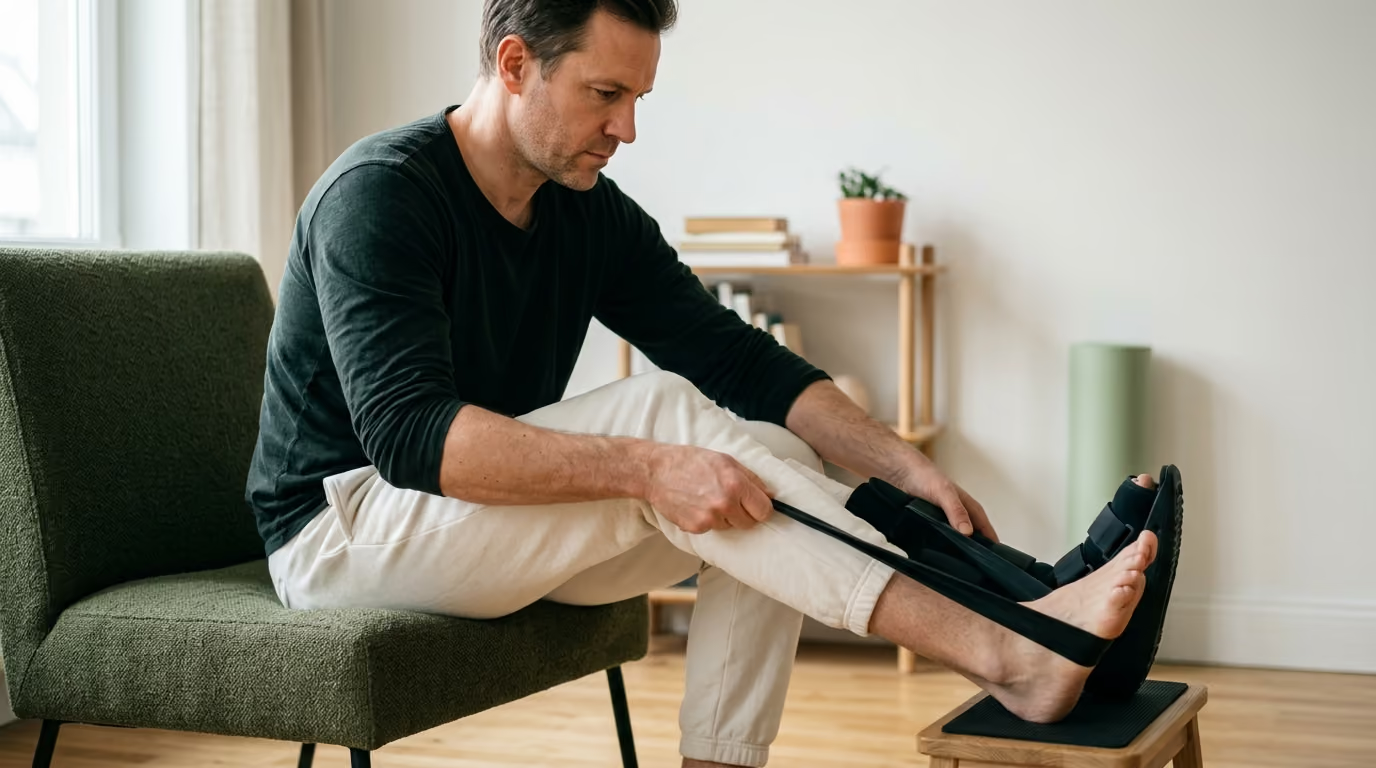
A patient recovering from open reduction and internal fixation (ORIF) of a stable bimalleolar or Weber B ankle fracture often follows an accelerated pathway. When surgical fixation is rigid and the surgical incisions have healed cleanly, the clinical team may permit early weight-bearing in a protective walking boot within two to three weeks.
Rehabilitation starts with gentle active dorsiflexion and plantarflexion out of the boot, progress to partial weight-bearing with crutches, and advances toward full weight-bearing as gait mechanics normalize. Strengthening focuses on rebuilding calf muscle volume, intrinsic foot strength, and ankle proprioception. Running and high-impact agility work are held until the patient demonstrates symmetrical single-leg calf raises, full ankle range of motion, and normal step-down mechanics.
Hip fracture management in older individuals focuses on immediate mobilization to prevent complications associated with prolonged bed rest. Surgical repairs, such as intramedullary nailing or hemiarthroplasty, are designed to allow immediate weight-bearing as tolerated.
Rehabilitation begins on the first post-operative day with assisted transfers, standing balance, and short walking bouts using a walker. Physical therapy focuses on hip abductor strengthening, quad activation, and fall prevention strategies. The long-term plan addresses home safety modifications, vision checks, balance training, and bone density management to reduce future fall and fracture risks.
Fractures that involve the weight-bearing surface of the knee joint require careful, measured loading. Even minor surface incongruities can accelerate joint degeneration under heavy compressive forces. As a result, these injuries often require strict non-weight-bearing protection for six to twelve weeks.
Rehabilitation during the non-weight-bearing phase emphasizes active and passive knee range of motion using continuous passive motion machines or guided physical therapy. Quad activation is maintained through straight-leg raises, neuromuscular electrical stimulation, and open-chain hip strengthening. Weight-bearing progresses slowly once imaging confirms articular consolidation, followed by extensive lower-chain strengthening and gait retraining.
Upper-extremity hand fractures follow different rehabilitation timelines than weight-bearing lower-limb breaks. Clinical literature on competitive athletes shows that return-to-sport timelines average roughly three weeks for metacarpal fractures and four weeks for phalangeal fractures when protected with custom splints or rigid casting.
Rehabilitation focuses on early tendon gliding exercises to prevent soft-tissue adhesions around the fracture site. Once stability is achieved, therapy emphasizes progressive grip strengthening, pinch grip development, wrist mobility, and fine motor coordination. Functional drills replicate the specific manual demands of the patient's trade or sport.
Patients with diabetes, severe peripheral neuropathy, or peripheral vascular disease require careful monitoring throughout rehabilitation. Blunted sensation increases the risk of skin breakdown, pressure sores from casts or braces, and unnoticed fixation failures.
Rehabilitation protocols for these individuals include daily skin inspections, careful offloading strategies, and extended healing timelines before advancing to full weight-bearing. Collaborative care involving endocrinologists, orthopedic surgeons, and wound care specialists helps protect both bone stability and soft-tissue health.
Progressing physical load after a fracture requires an objective, step-by-step process. Using a clear functional testing battery ensures that each step forward matches your current physical capacity.
Transitioning from an assistive device to unassisted walking should follow a gradual progression rather than an abrupt shift:
Before advancing to running, heavy strength training, or demanding occupational tasks, your physical performance should meet specific functional benchmarks:
Returning to high-impact activities like running, jumping, and tactical training follows a structured, graduated loading ladder:
Track your physical response after every progression. Mild muscular soreness is a normal adaptation to new physical demands, but sharp bone tenderness, escalating joint swelling, or an altered gait indicate that the mechanical load has exceeded current tissue capacity. When symptoms arise, reduce the volume or intensity to the previous successful step for several days before attempting to advance again.
While everyday muscular soreness and joint stiffness are normal parts of the recovery process, certain clinical symptoms point to serious complications like infection, deep vein thrombosis, compartment syndrome, or hardware failure.
Contact your orthopedic surgeon or seek urgent medical evaluation if you develop any of the following symptoms:
Prompt assessment of these warning signs protects your recovery, helps prevent long-term complications, and ensures that rehabilitation proceeds safely.
This guide is for educational and informational purposes only and does not constitute individual medical advice, diagnosis, or treatment. Fracture patterns, surgical procedures, and healing rates vary significantly between individuals. Always consult your orthopedic surgeon, physical therapist, or qualified healthcare professional before altering your weight-bearing status, stopping protective bracing, or advancing your physical rehabilitation program.
Post-traumatic swelling often persists long after biological bone union occurs. Inactivity, cast immobilization, and soft-tissue injury temporarily impair the local lymphatic and venous drainage systems. As you increase walking volume and daily standing, fluid naturally pools in the dependent extremity. Elevating the limb, using graduated compression garments if approved by your physician, and performing active muscle contractions will gradually restore normal circulation.
Yes. Muscle atrophy and joint stiffness are predictable biological adaptations to immobilization. When a joint is held static, the surrounding capsule tightens and connective tissues lose flexibility. At the same time, disuse leads to a rapid loss of muscle cross-sectional area and neuromuscular efficiency. Structured physical therapy focused on active range of motion, soft-tissue mobilization, and progressive resistance training will systematically rebuild joint mobility and muscular strength.
The effect of nonsteroidal anti-inflammatory drugs on bone healing remains a debated topic in orthopedic research. Some laboratory and clinical studies suggest that high-dose, long-term NSAID use may inhibit the inflammatory cascade necessary for early bone callus formation. Acetaminophen is often recommended for initial pain management when medically appropriate. Always discuss all pain medications with your treating physician to select the safest option for your specific injury.
Returning to driving depends on the location of the fracture, which limb was injured, whether you require a cast or walking boot, and whether you are taking impairing pain medications. For right-leg injuries, you must have adequate braking reaction time, normal muscle control, and full unassisted weight-bearing capacity before operating a vehicle safely. Always obtain formal clearance from your treating physician and consult your motor vehicle authority before resuming driving.
Follow BattleVet for practical guidance on military and veteran health, strength, recovery, testosterone, sleep and healthy aging. Stay connected for new articles, research backed insights and clear information to help you stay capable for the years ahead.


Build better habits around strength, recovery, sleep, hormones and healthy aging with practical guidance for active military personnel and veterans.
Explore BattleVet